1550nm非剥脱点阵激光脉冲时序调节成纤维细胞
2026年9月10日28 min read
Clinical, Cosmetic and Investigational Dermatology
PurposeNonablative fractional laser (NAFL) therapy is a widely used energy-based device (EBD) modality for dermal remodeling and photorejuvenation. Pulse temporal structure—the combination of peak power and pulse duration at constant total fluence—represents an underexplored determinant of cellular response. Type-N (normal mode) and Type-T (thermal mode) pulse configurations of a 1550-nm NAFL system induce distinct pathway-level transcriptional responses in human dermal fibroblasts. The fibroblast subpopulation-level correlates and functional tissue consequences of these differences remain unknown.
MethodsBulk RNA sequencing (n = 3 per group) was performed on human dermal fibroblasts at three days post-irradiation. Transcriptome-wide correlation scores were calculated against four reference fibroblast subpopulation clusters: secretory-reticular (cluster 1), pro-inflammatory (cluster 2), secretory-papillary (cluster 3), and mesenchymal (cluster 9). Subpopulation marker gene expression and collagen family profiles were visualized using z-score-normalized heat maps. Bioprinted three-dimensional (3D) dermal constructs were irradiated in Type-N or Type-T mode under matched parameters and assessed by uniaxial compression testing on Day 8.
ResultsControl fibroblasts showed transcriptional resemblance to the secretory-reticular subpopulation, consistent with a stable homeostatic baseline. Type-N mode–irradiated fibroblasts showed a broad, non-specific transcriptional activation pattern with signals consistent with pro-inflammatory, mesenchymal, and secretory-reticular subpopulation programs, without a single dominant subpopulation tendency, accompanied by broad induction across all collagen structural classes and, most prominently, fibrillar collagens. Type-T mode–irradiated fibroblasts showed preferential transcriptional resemblance to the pro-inflammatory subpopulation, consistently supported across replicates and marker gene analysis, with overall attenuation of fibrillar collagen expression and selective enrichment of basement membrane–associated collagens. Type-N mode–irradiated constructs showed directionally higher compressive modulus than Type-T mode and controls.
ConclusionPulse temporal structure is associated with distinct transcriptional tendencies in human dermal fibroblasts at equivalent total fluence. Type-T mode irradiation was associated with a robustly supported pro-inflammatory transcriptional tendency, while Type-N mode irradiation engaged a broad, non-specific activation pattern without preferential enrichment of a single subpopulation state. These molecular differences are directionally reflected in early biomechanical responses in the 3D dermal model and provide a framework for the mechanistic optimization of NAFL pulse parameters.
Keywords: non-ablative fractional laser、pulse structure、fibroblast heterogeneity、bulk RNA sequencing、bioprinted dermis
Nonablative fractional laser (NAFL) therapy is one of the most widely used energy-based device (EBD) modalities in dermatology and aesthetic medicine. By generating arrays of microscopic treatment zones (MTZs) within the dermis while preserving the overlying epidermis, NAFL devices initiate a controlled wound-healing response that drives neocollagenesis, dermal matrix remodeling, and restoration of the dermal–epidermal junction (DEJ). 1 , 2 Unlike ablative fractional systems, the non-ablative approach minimizes post-procedural downtime and reduces the risk of adverse events, making it suitable for a broad range of treatment indications, including photoaging, acne, surgical scarring, and pigmentary disorders. 3 , 4 The 1550-nm erbium-glass wavelength, in particular, is primarily absorbed by water and can deliver controlled photothermal energy into the dermis with relatively limited melanin absorption, supporting its application across diverse skin phototypes. 5 , 6
Accumulating evidence suggests that the biological effects of NAFL extend beyond simple stimulation of fibroblast activity. Dermal remodeling after nonablative phototherapy involves complex cascades, including regulation of matrix metalloproteinases, activation of heat-shock responses, modulation of growth factor signaling, and remodeling of DEJ, all of which are influenced by the biophysical characteristics of the delivered energy. 4 A central question in this context is whether different pulse configurations are associated with the preferential enrichment of distinct fibroblast subpopulation-level transcriptional states. Human dermal fibroblasts are functionally and spatially heterogeneous, comprising at least four transcriptionally discrete subpopulations identified by single-cell RNA sequencing: secretory-reticular, pro-inflammatory, secretory-papillary, and mesenchymal. 7 Secretory-papillary fibroblasts reside predominantly in the papillary dermis adjacent to the DEJ, where they contribute to both structural matrix organization and keratinocyte-fibroblast crosstalk. In contrast, pro-inflammatory fibroblasts are characterized by chemokine-driven signaling programs and high expression of APOE, a key mediator of lipid transport and cholesterol homeostasis. 7 , 8 Identifying which subpopulation-level programs are preferentially engaged by different laser pulse structures may provide mechanistic insight into differences in the observed pathway-level programs and inform rational parameter selection for targeted clinical outcomes.
Beyond transcriptomic profiling, a critical translational question is whether pulse structure–dependent molecular differences are reflected at the functional tissue level. To address this, we employed a bioprinted three-dimensional (3D) dermal model that preserved the volumetric scaffold architecture and enabled the quantitative biomechanical assessment following laser treatment. 9 The compressive mechanical properties of the dermis are largely determined by its fibrillar collagen network, 10 , 11 making compression testing a relevant functional readout for ECM remodeling transcriptional programs. In this study, we integrated transcriptome-wide cluster correlation scoring, subpopulation marker gene analysis, collagen family profiling, and 3D construct compression testing to provide a multilevel characterization of how the NAFL pulse structure shapes fibroblast biology and dermal biomechanics.
Human dermal fibroblasts (Lonza, Basel, Switzerland) were maintained in Dulbecco’s modified Eagle’s medium (DMEM; Gibco, Thermo Fisher Scientific, Waltham, MA, USA) supplemented with 10% fetal bovine serum (FBS; Gibco) and 1% penicillin-streptomycin (Gibco) at 37°C in a humidified atmosphere containing 5% CO 2 . All experiments were performed using cells within a restricted passage range to minimize phenotypic drift. Irradiation was performed using a 1550-nm NAFL system (MOSAIC 3D; Cynosure Lutronic Inc., Goyang, Republic of Korea) under two configurations: Type-N (normal mode: higher peak power, shorter pulse duration) and Type-T (thermal mode: lower peak power, longer pulse duration), both delivering equivalent total energy per unit area. The laser parameters were adjusted so that the total delivered energy was comparable between the two modes, whereas peak power and pulse duration differed (spot density: 500 spots/cm 2 ; fluence: 5 mJ/cm 2 ). Untreated cells served as controls. Cells were returned to standard culture conditions immediately after irradiation and were harvested three days post-treatment.
Total RNA was extracted using standard column-based protocols, and quality was assessed by spectrophotometric and electrophoretic methods before library preparation. Strand-specific RNA-sequencing libraries were prepared using the TruSeq Stranded Total RNA Library Prep Gold Kit (Illumina, San Diego, CA, USA) and sequenced by Macrogen Inc (Seoul, Republic of Korea) using paired-end reads (2 × 101 bp), generating approximately 7.0 Gb per sample. Three biological replicates were included for each experimental group (n = 3). Raw FASTQ reads were quality-assessed using FastQC (v.0.11.5) and aligned to the human reference genome (GRCh38) using STAR (v.2.7.11b) with Ensembl release 112 gene annotations. Transcript-level quantification was performed using RSEM (v.1.3.1). Differential gene expression analysis was conducted using DESeq2 (v.1.48.1) with apeglm log2 fold-change shrinkage. Raw read counts were normalized to transcripts per million (TPM) using gene length information. TPM values were calculated using all annotated genes with available gene length information (n = 63,240 genes).
Subpopulation-level analysis was performed using two complementary approaches, considering that bulk RNA sequencing of a monoculture cannot resolve individual cell identities but can detect transcriptional tendencies consistent with known subpopulation programs. First, transcriptome-wide Surrogate Proportion Variable (SPV) scores were calculated between each bulk RNA-seq sample and the four fibroblast subpopulation reference clusters identified in the human skin single-cell RNA sequencing atlas of Solé-Boldo et al 7 secretory-reticular (Cluster 1), pro-inflammatory (Cluster 2), secretory-papillary (Cluster 3), and mesenchymal (Cluster 9). The mean correlation scores per experimental group were used to quantitatively rank subpopulation similarities. Second, z-score-normalized expression of representative subpopulation marker genes was visualized as a heatmap: secretory-reticular markers ( WISP2/CCN5, SLPI, CTHRC1, MFAP5, TSPAN8 ), pro-inflammatory markers ( CCL19, APOE, CXCL2, CXCL3, EFEMP1 ), secretory-papillary markers ( APCDD1, ID1, WIF1, COL18A1, PTGDS ), and mesenchymal markers ( ASPN, POSTN, GPC3, TNN, SFRP1 ). 7 Results are interpreted as transcriptional tendencies rather than definitive cell type classifications.
Normalized expression values (transcripts per million) were extracted for all detected collagen family members. Z-score-normalized expression across samples was visualized as a heatmap. Collagen genes were annotated according to the established structural classification: 10 , 12 fibrillar collagens ( COL1, COL2, COL3, COL5, and COL11 ), basement membrane/network collagens ( COL4, COL8, and COL10 ), fibril-associated collagens with interrupted triple helices ( COL9, COL12, COL14, COL16, COL19, COL20, and COL21 ), transmembrane collagens ( COL13, COL17, COL23, and COL25 ), and multiplexin collagens ( COL15 and COL18 ). Group-level mean z-scores were calculated for each structural class to enable a quantitative comparison across conditions.
Bioprinted three-dimensional dermal constructs were fabricated by collagen microextrusion seeded with human dermal fibroblasts, as previously described. 11 Briefly, constructs were maintained under liquid–liquid interface (LLI) culture conditions for 7–14 days to allow fibroblast-mediated matrix compaction and maturation prior to laser treatment. Constructs were irradiated in Type-N or Type-T mode at 4 mJ per microzone with equivalent spot density settings. Untreated constructs served as controls.
The compressive mechanical properties of the 3D dermal constructs were assessed on day 8 post-irradiation using uniaxial compression testing. Constructs were placed between parallel plates and compressed at a controlled rate. Stress–strain curves were recorded, and the compressive modulus was determined from the slope of the linear elastic region (R 2 > 0.95 for all included measurements). Data are presented as mean ± standard deviation (SD) (n = 3 per group).
All transcriptomic analyses were performed using R (v.4.5.0). Statistical significance for differential gene expression was defined as FDR-adjusted p < 0.1 with apeglm-shrunk log2 fold-change estimates. Biomechanical data are summarized as mean ± standard deviation.
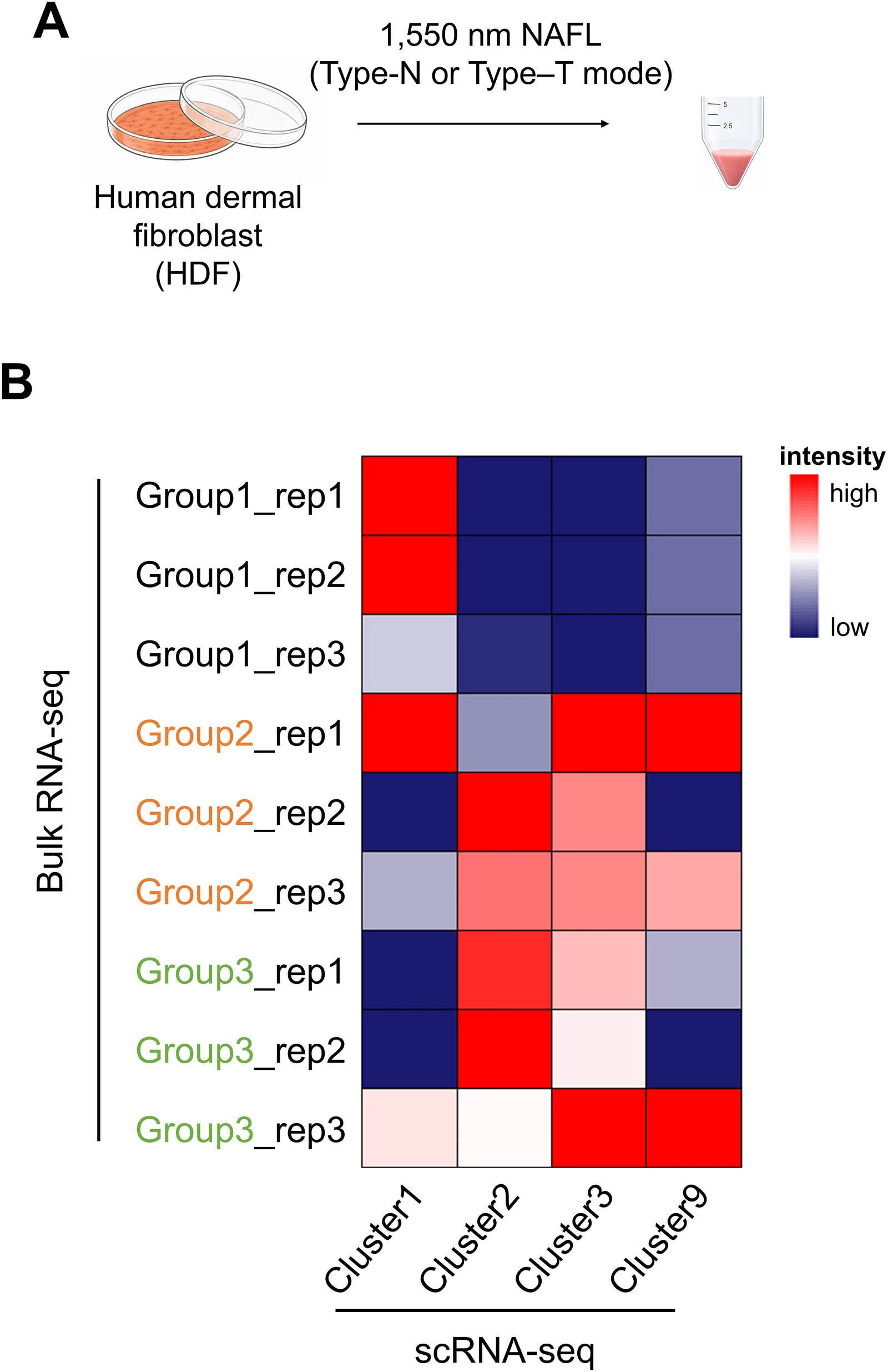
Transcriptome-wide similarity scores against four reference fibroblast subpopulation clusters from Solé-Boldo et al 7 revealed consistent group-level differences in subpopulation similarities ( Figure 1 ). For each group, all four cluster scores are reported in descending order to provide a full ranking of subpopulation resemblance, rather than identifying a single dominant tendency. Control fibroblasts (Group 1) showed the highest SPV score for Cluster 1 (secretory-reticular; +0.283), followed by Cluster 9 (mesenchymal; −0.155), Cluster 2 (pro-inflammatory; −0.374), and Cluster 3 (secretory-papillary; −0.402). Only Cluster 1 showed a positive SPV score, and all three replicates independently ranked Cluster 1 first (Group1_rep1: +0.582; Group1_rep2: +0.325; Group1_rep3: −0.058), with the lowest inter-replicate variability of all three groups (mean SD across clusters = 0.116). This pattern is consistent with a stable, homeostatic baseline state in which secretory-reticular transcriptional programming predominates.
Type-N mode–irradiated fibroblasts (Group 2) showed the following relative cell-type abundance cluster score ranking: Cluster 3 (secretory-papillary; +0.295), Cluster 9 (mesenchymal; +0.173), Cluster 2 (pro-inflammatory; +0.125), and Cluster 1 (secretory-reticular; −0.074). Notably, the three highest-ranking clusters all yielded SPV scores. However, inter-replicate variability in Group 2 was substantially higher than in the other groups (mean SD = 0.313 versus 0.116 in Group 1 and 0.175 in Group 3), indicating that the group-mean ranking may not represent a stable biological tendency. Examination of individual replicates revealed markedly divergent patterns: Group2_rep1 ranked Cluster 9 (mesenchymal) highest (+0.801), followed by Cluster 3 (+0.648), whereas Group2_rep2 and Group2_rep3 both ranked Cluster 2 (pro-inflammatory) highest (+0.354 and +0.140, respectively). Considered together, the similarity analysis does not converge on a single dominant subpopulation. Rather, transcriptional signals consistent with pro-inflammatory (supported by two of three replicates at the individual level), mesenchymal, and, to a lesser extent, secretory-papillary programs were all detectable within this group. Type-T mode–irradiated fibroblasts (Group 3) showed the following positive SPV score ranking: Cluster 2 (pro-inflammatory; +0.248), Cluster 3 (secretory-papillary; +0.108), Cluster 9 (mesenchymal; −0.019), and Cluster 1 (secretory-reticular; −0.209). The pro-inflammatory cluster was ranked first in two of three replicates (Group3_rep1: +0.207; Group3_rep2: +0.534). The third replicate (Group3_rep3) showed a more distributed pattern with Cluster 9 (mesenchymal; +0.274) and Cluster 3 (secretory-papillary; +0.246) narrowly leading, although Cluster 2 also remained positive (+0.003). The secondary tendency toward secretory-papillary (group mean +0.108) was notable, though substantially lower than the primary pro-inflammatory score. Taken together, the pro-inflammatory tendency in Group 3 was more robustly supported across replicates than any single tendency in Group 2. Quantitative scores and subpopulation rankings are summarized in Table 1 .
Table 1 Quantitative Subpopulation SPV Scores and Transcriptional Tendencies Across Experimental Groups
| Group | Condition | Closest Cluster (Mean Score) | Subpopulation Tendency | Key Transcriptomic Features |
|---|---|---|---|---|
| 1 | Control | Cluster 1 (+0.283) > Cluster 9 (−0.155) > Cluster 2 (−0.374) | Secretory-reticular–like (1°); all others negative | Homeostatic baseline; no dominant activation program |
| 2 | Normal mode (Type-N) | Cluster 3 (+0.295) > Cluster 9 (+0.173) > Cluster 2 (+0.125) [SD=0.313; high variability] | Multi-subtype signals (Pro-inflammatory by 2/3 replicates in SPV; Mesenchymal ≈ Secretory-reticular by z-score); no single dominant tendency | Broad fibrillar collagen induction (COL1/3/5/6); ECM organization pathways; signals from pro-inflammatory (SPV, 2/3 reps), mesenchymal, and secretory-reticular programs all present; high inter-replicate variability (SD = 0.313) |
| 3 | Thermal mode (Type-T) | Cluster 2 (+0.248) > Cluster 3 (+0.108) > Cluster 9 (−0.019) | Pro-inflammatory–like (1°); Secretory-papillary–like (2°) | Lipid/SREBP metabolic reprogramming; selective BM-associated collagen enrichment (COL8A2, COL10A1, COL18A1); secondary Secretory-papillary tendency (z-score 2°: +0.319; marker consistency 3/5) |
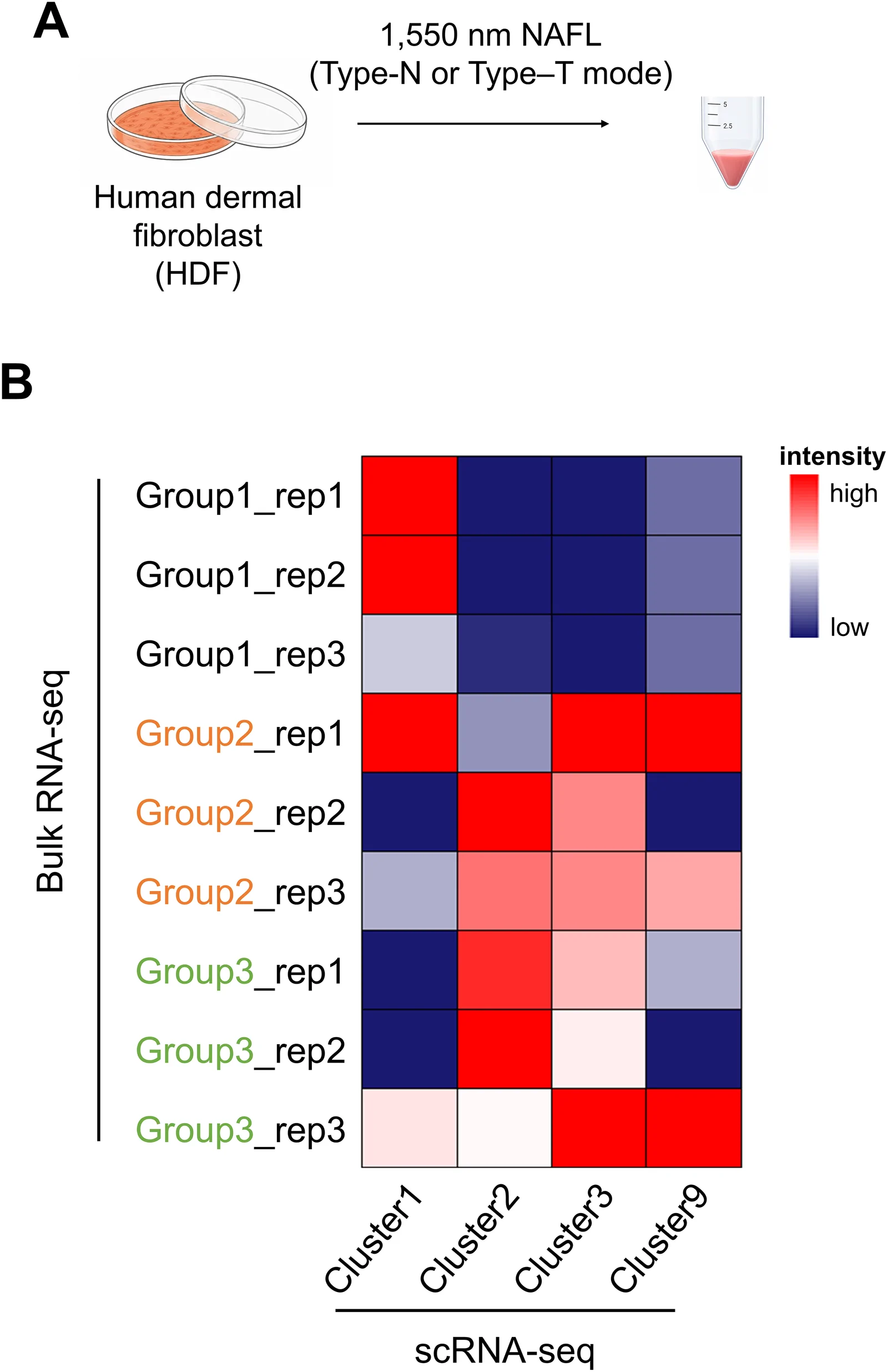
Figure 1 Transcriptome-wide cluster SPV analysis of bulk RNA-seq profiles against human dermal fibroblast subpopulation reference signatures. (A) Schematic of experimental design. Human dermal fibroblasts (HDF) were irradiated with a 1550-nm NAFL system (MOSAIC 3D) in Type-N (normal mode) or Type-T (thermal mode) at equivalent total fluence, with untreated cells serving as controls. Cells were harvested at day 3 post-irradiation for RNA sequencing. (B) Heatmap depicting SPV scores between each bulk RNA-seq sample (rows; Group 1 = control [unlabeled], Group 2 = Type-N mode [Orange], Group 3 = Type-T mode [green]; n = 3 per group) and four reference fibroblast subpopulation cluster gene signatures (columns; Cluster 1 = secretory-reticular, Cluster 2 = pro-inflammatory, Cluster 3 = secretory-papillary, Cluster 9 = mesenchymal) as defined by Solé-Boldo et al7 Color intensity represents the SPV score (red = high positive correlation, blue = low/negative correlation). Group mean scores: Group 1/Cluster 1 = +0.283; Group 2/Cluster 3 = +0.295; Group 3/Cluster 2 = +0.248. Scores reflect transcriptional resemblance to reference subpopulation programs rather than definitive cell identity classification.转录组学簇 SPV 分析:bulk RNA-seq 谱与 4 类成纤维细胞亚群参考特征的相似性评分(含实验设计示意)。
Heatmap visualization of 20 representative subpopulation marker genes across all nine samples provided complementary, but partially discordant, evidence relative to the SPV scores ( Figure 2 ). Z-score–based ranking of subpopulation marker sets for each group is reported below alongside the Pearson r rankings to enable direct comparison. Control fibroblasts (Group 1) showed the lowest mean z-scores across all four marker sets (secretory-reticular: −0.125; mesenchymal: −0.219; pro-inflammatory: −0.268; secretory-papillary: −0.276), with all scores negative. Despite the small absolute differences between ranks, the secretory-reticular set showed the least negative mean value, consistent with the Pearson r result. This pattern reflects the quiescent homeostatic baseline expected in unirradiated fibroblasts.
Type-N mode–irradiated fibroblasts (Group 2) showed the following z-score marker ranking: mesenchymal (+0.119; marker consistency 2/4), secretory-reticular (+0.072; 1/5), secretory-papillary (−0.043; 1/5), and pro-inflammatory (−0.124; 0/5). The difference between the first-ranked (mesenchymal, +0.119) and second-ranked (secretory-reticular, +0.072) marker sets was only +0.047, a margin that is insufficient to distinguish the two subpopulations with confidence. Among individual markers, POSTN and SFRP1 were the most consistently elevated mesenchymal markers, while SLPI and CTHRC1 showed elevated expression among secretory-reticular markers in individual samples. Taken together, the z-score analysis converges with the Pearson r analysis in demonstrating that Type-N mode–irradiated fibroblasts do not exhibit a single dominant subpopulation tendency. Transcriptional signals consistent with pro-inflammatory (supported by two of three replicates in Pearson r analysis), mesenchymal, and secretory-reticular programs were all present, while pro-inflammatory markers showed the lowest z-score mean (−0.124; marker consistency 0/5), indicating that pro-inflammatory was the most discordant between the two methods. The data as a whole are most consistent with a broad, non-specific transcriptional activation pattern that partially engages multiple subpopulation programs rather than preferential engagement of a single defined subpopulation state.
Type-T mode–irradiated fibroblasts (Group 3) showed the following z-score marker ranking: pro-inflammatory (+0.391; marker consistency 4/5), secretory-papillary (+0.319; 3/5), mesenchymal (+0.101; 1/4), and secretory-reticular (+0.054; 2/5). This ranking is broadly consistent with the SPV result and provides the strongest cross-method concordance of the three groups. Pro-inflammatory markers including CCL19, APOE, CXCL2 , and CXCL3 , were elevated in two of the three replicates, with the third showing a more distributed pattern.
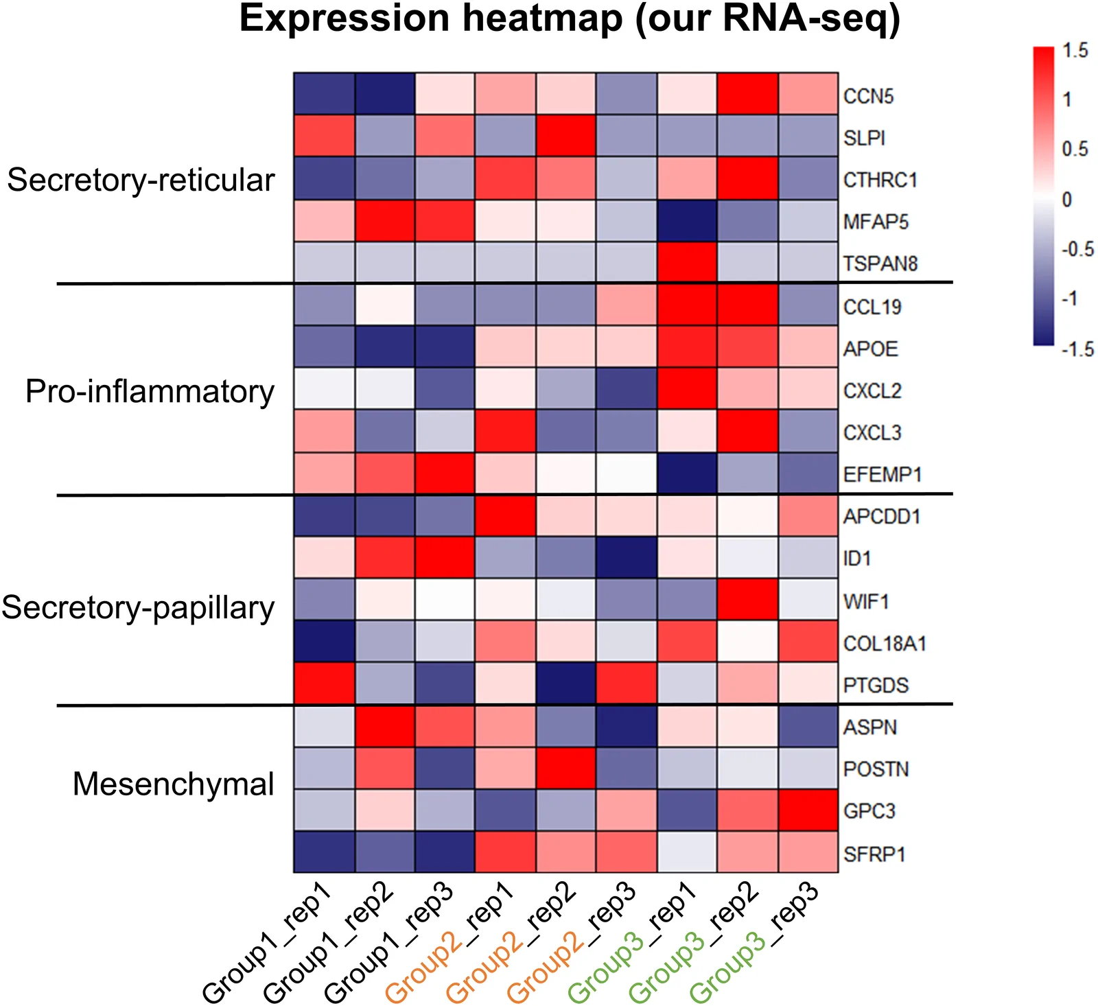
Figure 2 Expression of fibroblast subpopulation marker genes across experimental groups. Heatmap depicting z-score-normalized expression values (scale: −1.5 to +1.5; red = high, blue = low) of 20 representative marker genes corresponding to four fibroblast subpopulations, secretory-reticular (CCN5/WISP2, SLPI, CTHRC1, MFAP5, TSPAN8), pro-inflammatory (CCL19, APOE, CXCL2, CXCL3, EFEMP1), secretory-papillary (APCDD1, ID1, WIF1, COL18A1, PTGDS), and mesenchymal (ASPN, POSTN, GPC3, SFRP1), across all nine samples (Group 1 [control, unlabeled], Group 2 [Type-N mode, Orange label], Group 3 [Type-T mode, green label]; n = 3 per group). Group 2 shows relative enrichment of secretory-papillary markers (APCDD1, ID1) in most replicates; Group 3 shows relative enrichment of pro-inflammatory markers (CCL19, APOE, CXCL2, CXCL3) in two of three replicates. Marker genes selected based on Solé-Boldo et al (2020).7 Results should be interpreted as transcriptional tendencies.各实验组成纤维细胞亚群标志基因表达热图(z-score 标准化)。
Collagen family profiling revealed clear quantitative differences in the extent and pattern of collagen gene expression between the two irradiation modes ( Figure 3 ).
Type N-mode-irradiated fibroblasts (group 2) had the highest mean z-scores across all four collagen structural classes: fibrillar collagens (+0.268), basement membrane/network collagens (+0.207), FACIT collagens (+0.211), and other structural collagens (+0.245). At the individual gene level, the most prominent fibrillar collagens included COL1A1 (group 2: +0.302 vs group 3: +0.195), COL1A2 (+0.638 vs +0.294), COL3A1 (+0.509 vs +0.262), COL5A1 (+0.735 vs +0.115), and COL6A1, COL6A2 , and COL6A3 (+0.610, +0.313, +0.572, +0.330, +0.483, and +0.194, respectively). This broad collagen induction pattern across all structural classes was consistent with active structural ECM remodeling.
Type-T mode–irradiated fibroblasts (Group 3) exhibited overall attenuation of fibrillar collagen expression relative to Type-N mode (mean fibrillar class z-score: −0.004 vs +0.268). However, three basement membrane- and interface-associated collagens showed selectively higher expression in Group 3 compared to Group 2: COL8A2 (+0.834 vs +0.244), COL10A1 (+0.379 vs −0.185), and COL18A1 (+0.731 vs +0.258). Notably, COL18A1 is a canonical marker of the secretory-papillary fibroblast subpopulation 8 and a structural component of the DEJ. Its selective enrichment in Type-T mode fibroblasts, within the context of overall fibrillar collagen attenuation, is consistent with a transcriptional state oriented toward DEJ-adjacent interface remodeling rather than bulk matrix deposition.
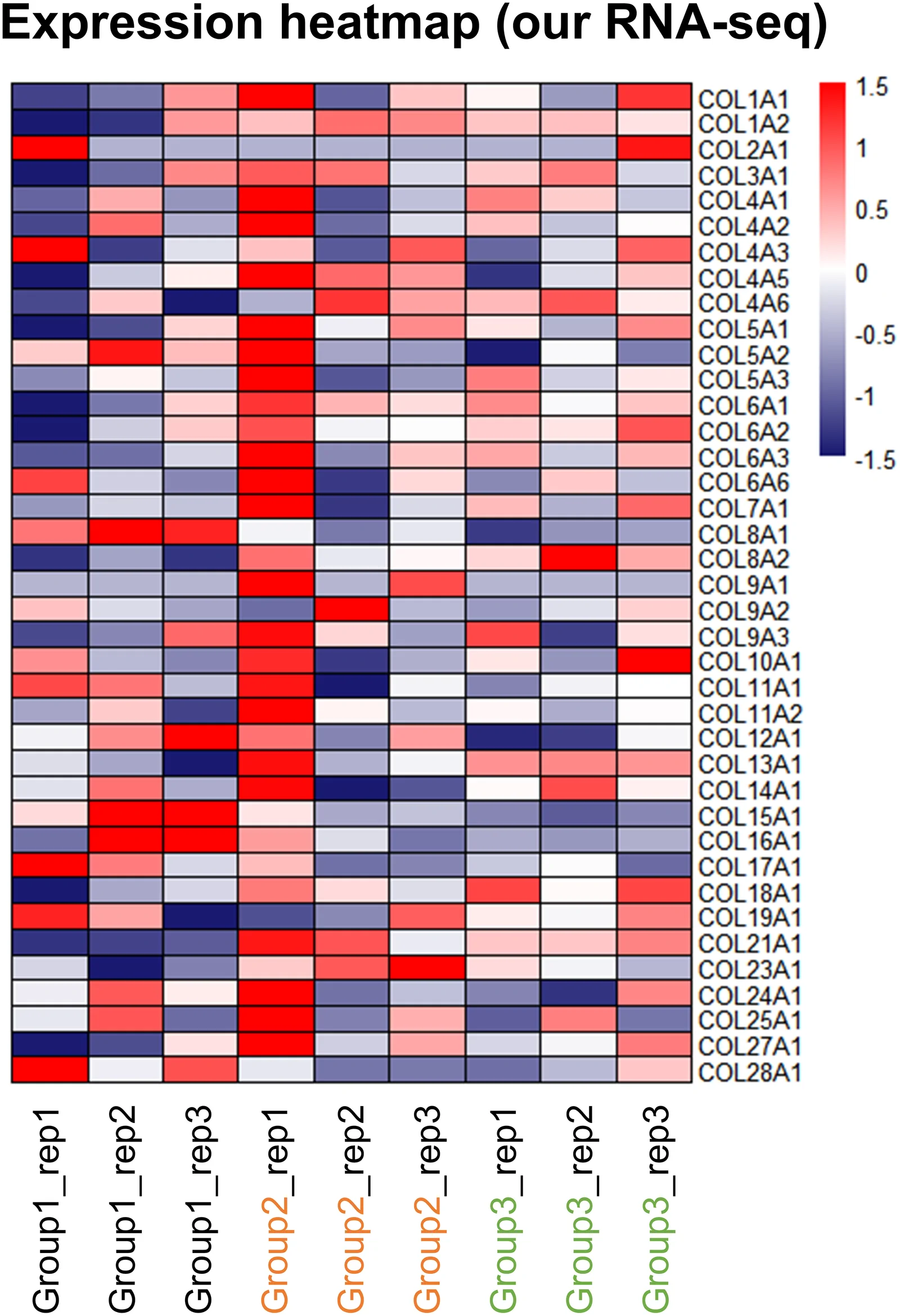
Figure 3 Collagen family expression profiles in Type-N mode and Type-T mode–irradiated fibroblasts. Heatmap depicting z-score-normalized expression values (scale: −1.5 to +1.5) of all detectable collagen family members across Group 1 (control, unlabeled), Group 2 (Type-N mode, Orange label), and Group 3 (Type-T mode, green label) samples (n = 3 per group). Group 2 shows broad collagen induction across all structural classes; group-level mean z-scores: fibrillar = +0.268, basement membrane/network = +0.207, FACIT = +0.211. Group 3 shows overall attenuation of fibrillar collagen expression (mean fibrillar z-score = −0.004) with selective enrichment of specific basement membrane–associated collagens: COL8A2 (+0.834 vs +0.244 in Group 2), COL10A1 (+0.379 vs −0.185), and COL18A1 (+0.731 vs +0.258).Type-N 与 Type-T 照射后成纤维细胞胶原家族表达谱热图。
To determine whether the transcriptional and collagen expression differences associated with the two pulse structures were reflected in functional tissue-level outcomes, we assessed the compressive mechanical properties of the bioprinted 3D dermal constructs irradiated with matched parameters ( Figure 4 ).
Figure 4A shows representative images of a bioprinted 3D dermal construct model fabricated using collagen microextrusion and fibroblast seeding under LLI culture conditions. Figure 4B and C illustrates the compression-testing procedure with pre- and post-compression images of representative constructs. Figure 4D presents the compressive modulus values on day 8 post-irradiation for control, Type-N, and Type-T conditions. Untreated control constructs exhibited a mean compressive modulus of 12.0 ± 4.8 kPa. Type-N mode-irradiated constructs exhibited a higher mean value of 25.5 ± 16.2 kPa, whereas Type-T mode-irradiated constructs exhibited an intermediate value of 18.1 ± 6.9 kPa.
The directional ordering (Type-N > Type-T > control) was consistent with the transcriptomic and collagen profiling data. Type-N mode, associated with a secretory-papillary-like transcriptional profile and broad fibrillar collagen induction across all structural classes, would be mechanistically expected to drive greater early matrix stiffening, as fibrillar collagens are the primary load-bearing determinants of dermal compressive resistance. 10 , 11 In contrast, Type-T mode, associated with a pro-inflammatory-like profile and overall attenuation of fibrillar collagen expression despite selective BM-associated collagen enrichment, would be expected to produce a more attenuated early mechanical response. These findings should be interpreted as directional, hypothesis-generating findings consistent with the molecular data.
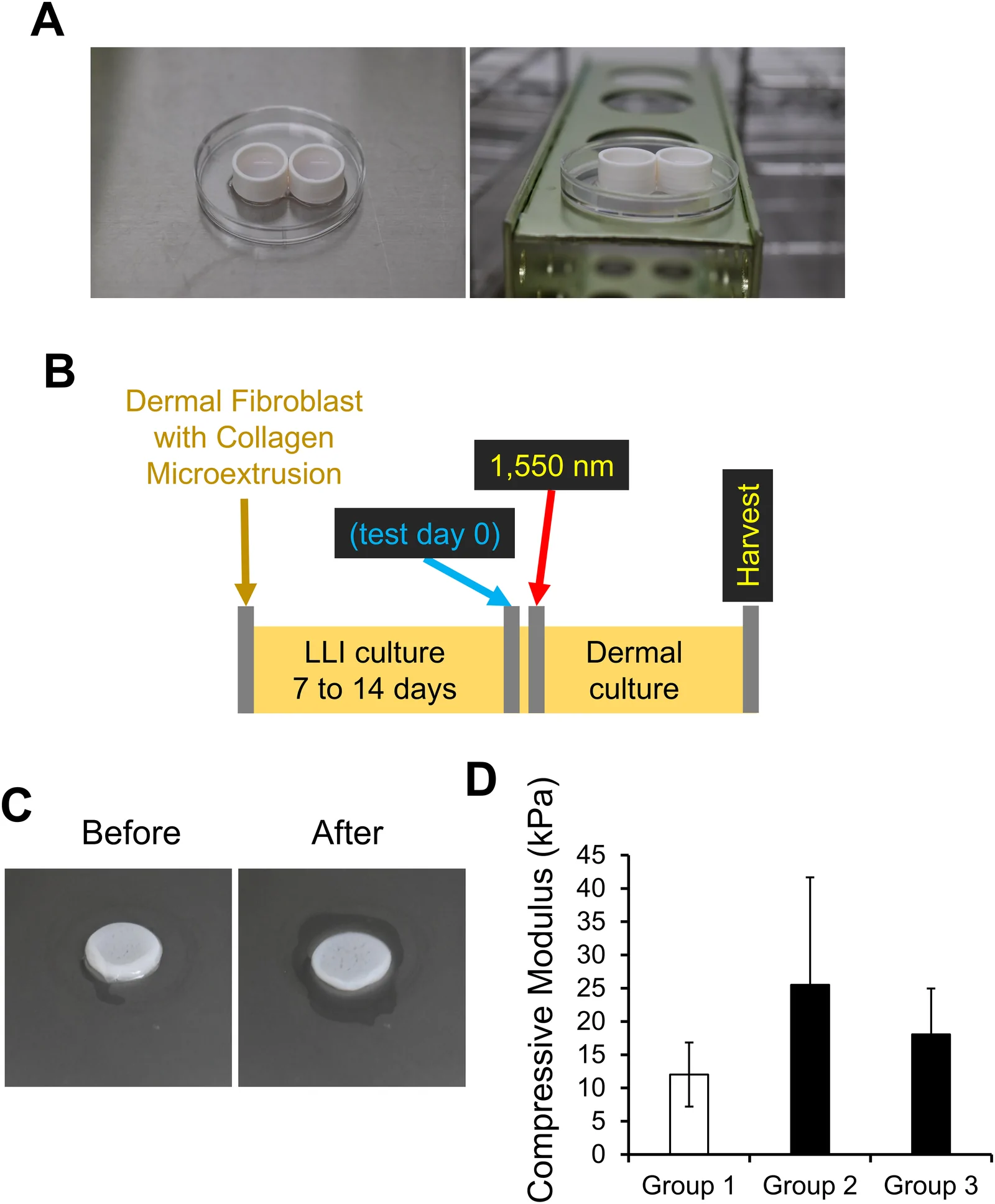
Figure 4 Bioprinted 3D dermal model and compressive mechanical response to pulse structure–specific irradiation. (A) Representative photographs of the bioprinted 3D dermal construct (collagen microextrusion with embedded human dermal fibroblasts) unDer Standard laboratory conditions. (B) Schematic timeline of construct preparation and irradiation: constructs were fabricated by collagen microextrusion and maintained under liquid–liquid interface (LLI) culture for 7–14 days to allow matrix compaction, irradiated on test day 0, and harvested for analysis at day 8. (C) Representative photographs of constructs before (left) and after (right) uniaxial compression testing are shown. (D) Bar graph showing mean compressive modulus (kPa) ± SD (n = 3) at day 8 post-irradiation for Group 1 (Control: 12.0 ± 4.8 kPa), Group 2 (Type-N mode at 4 mJ: 25.5 ± 16.2 kPa), and Group 3 (Type-T mode at 4 mJ: 18.1 ± 6.9 kPa). Results are presented as directional, exploratory findings consistent with transcriptomic data.生物打印 3D 真皮模型及不同脉冲结构照射后的压缩力学响应。
This study provides the first multilevel characterization of how the temporal structure of NAFL pulses shapes fibroblast biology beyond the pathway level by integrating transcriptome-wide subpopulation correlation scoring, marker gene analysis, collagen family profiling, and functional 3D biomechanics. The principal finding is that at equivalent total fluence, Type-N and Type-T irradiation are associated with transcriptional tendencies that most resemble the secretory-papillary and pro-inflammatory fibroblast subpopulations, respectively, as defined by the human skin single-cell atlas. 7 Importantly, these molecular differences are directionally reflected in the early compressive mechanical response of a bioprinted 3D dermal model, providing preliminary functional evidence that pulse structure-dependent transcriptional prioritization has measurable tissue-level consequences. These findings are broadly consistent with the multifactorial nature of skin regeneration, in which fibroblast activation, collagen synthesis, extracellular matrix remodeling, inflammatory modulation, and growth factor signaling collectively govern tissue repair outcomes. 13
The transcriptional profile of Type-N mode–irradiated fibroblasts warrants careful interpretation in the context of known fibroblast subpopulation biology. As detailed in the Results, neither the transcriptome-wide SPV analysis nor the z-score marker gene analysis yielded a single dominant subpopulation assignment for Group 2. The SPV analysis identified signals consistent with pro-inflammatory (supported in two of three individual replicates), mesenchymal, and secretory-papillary programs, while the z-score marker analysis showed mesenchymal and secretory-reticular programs at comparable levels, with pro-inflammatory markers showing the lowest mean and zero marker consistency. Taken across both analytical approaches, the most parsimonious interpretation is that Type-N mode–irradiated fibroblasts exhibited transcriptional signals broadly consistent with pro-inflammatory, mesenchymal, and secretory-reticular subpopulation programs simultaneously, without preferential enrichment of any single state. This pattern is consistent with a broad, non-specific ECM remodeling response rather than a directed subpopulation shift. With this caveat, the broad collagen induction observed in Type-N mode–irradiated fibroblasts—spanning fibrillar (COL1/3/5), network (COL4A5), and FACIT collagen families—is consistent with an active structural matrix-building program. Both mesenchymal and secretory-papillary fibroblasts are known to contribute to structural ECM organization, and the observed collagen induction pattern is compatible with activation of programs shared across these subpopulation states. Furthermore, the directional association between this transcriptional–collagen profile and higher early compressive modulus in the 3D model is consistent with the established role of fibrillar collagens, particularly types I and III, as primary load-bearing determinants of dermal mechanical stiffness. 10 , 14 , 15
The pro-inflammatory tendency of the type-T mode-irradiated fibroblasts, together with the lipid metabolic pathway enrichment identified previously, suggests a mechanistically coherent transcriptional program. Pro-inflammatory fibroblasts are characterized by high expression of APOE , 7 a key regulator of lipid transport, cholesterol efflux, and lipoprotein homeostasis. The SREBF1/INSIG1/PPARA/DHCR7/SCD lipid biosynthesis program, activated in type-T mode-irradiated cells, operates within the same metabolic space as APOE-mediated lipid handling and may reflect an integrated inflammatory–metabolic program characteristic of the pro-inflammatory fibroblast transcriptional state. This interpretation is further supported by the known intersection of prostaglandin, eicosanoid, and cholesterol pathways in inflammatory stromal signaling. 16 The selective enrichment of COL18A1 , which encodes both a structural basement membrane component and endostatin—a bioactive fragment with angiogenic and anti-inflammatory properties—together with overall fibrillar collagen attenuation in Type-T mode fibroblasts, suggests a remodeling program oriented toward maintenance of the DEJ interface rather than bulk dermal matrix reinforcement.
From a translational perspective, the distinct transcriptional tendencies associated with each pulse mode may have practical implications for parameter selection in clinical NAFL practice. The secretory-papillary–like, broad ECM-remodeling program of the Type-N mode—manifested as fibrillar collagen induction and directionally higher early matrix stiffness—may be particularly relevant for indications requiring structural dermal rejuvenation, such as rhytide reduction, laxity, and atrophic scarring. 3 , 4 In contrast, the pro-inflammatory–like, lipid-metabolic program associated with Type-T mode may be more relevant for conditions involving dermal–epidermal interface dysregulation, such as melasma, in which disruption of the DEJ basement membrane and inflammatory signaling in the papillary dermis are increasingly recognized as key pathological drivers. 17 , 18 Fibroblast-to-keratinocyte communication via paracrine signaling and extracellular vesicles represents a potential mechanism by which the pro-inflammatory transcriptional state of Type-T mode fibroblasts could influence epidermal pigmentary regulation, 19–21 although this hypothesis requires direct experimental validation. These translational applications remain speculative at this stage and will require validation through appropriate preclinical and clinical studies before informing clinical practice.
This study had several important limitations that should be acknowledged. First, the inference of fibroblast subpopulation tendencies from bulk RNA sequencing of monocultures was inherently indirect. The observed correlation scores and marker gene patterns reflect transcriptional tendencies within a uniform fibroblast population rather than true changes in subpopulation composition. More specific validation at the transcript and protein level, such as quantitative PCR and protein-based assays, will be required to substantiate these findings. Direct single-cell sequencing of laser-irradiated fibroblasts is required to definitively resolve cell-state heterogeneity. Second, the compression testing data were preliminary (n = 3) with substantial inter-construct variability; statistical validation requires larger sample sizes and potentially extended time points to capture the full trajectory of laser-induced matrix remodeling. These biomechanical findings will need to be further substantiated through histological and ultrastructural evidence of collagen organization and extracellular matrix deposition. Third, all experiments were conducted in vitro or in bioprinted constructs, and translation to in vivo dermal biology requires validation using appropriate preclinical or clinical models. Fourth, protein-level, lipidomic, and extracellular vesicle profiling data are needed to assess the downstream functional consequences of the observed transcriptional programs.
Despite these limitations, the multilevel consistency of our findings—from transcriptome-wide correlation scores to marker gene expression, collagen family patterns, and biomechanical behavior—provides comprehensive converging evidence that the pulse temporal structure shapes fibroblast biology in a biologically meaningful manner. These findings should encourage prospective studies with larger sample sizes, in vivo validation, and correlation with clinical outcomes to determine whether pulse mode selection can be systematically aligned with patient-specific therapeutic objectives.
This study demonstrated that pulse temporal structure, independent of total fluence, was a biologically active variable that shaped fibroblast transcriptional identity and downstream tissue biomechanics. Type-N mode irradiation engaged a broad, ECM-remodeling–oriented transcriptional program associated with greater early matrix stiffness, whereas Type-T mode irradiation preferentially engaged a pro-inflammatory, lipid-metabolic program associated with basement membrane–related collagen remodeling. In short, altering the temporal delivery of laser energy—not just its total dose—directed fibroblasts toward distinct functional states, offering a mechanistic basis for tailoring NAFL pulse parameters to specific clinical indications. These findings should be further validated through larger-scale, in vivo, and clinical studies.
循证层面:本研究为体外(细胞 + 3D 构建体)机制研究,证据等级为基础/转化层面,尚不能直接外推至临床疗效;但首次在亚群水平上阐明脉冲时序结构是独立于总能量密度的生物活性变量。
局限:样本量小(每组 n=3)、仅观察照射后 3 天转录与第 8 天力学,缺乏长期、在体与临床终点验证;Type-N"广泛激活"与 Type-T"促炎倾向"的功能后果需更大规模研究确认。
临床提示:在设备参数设置时,除关注总能量密度/脉宽绝对值外,可考虑以脉冲时序(峰值功率×脉宽)作为调节真皮重塑方向的抓手:偏向 ECM/纤维胶原重塑(Type-N 特征)或偏向炎症/基底膜胶原重塑(Type-T 特征)可对应不同适应证的差异化需求;上述结论需以临床参数研究与随机对照试验进一步验证后再行落地。
声明:中文精读 · 仅供学术参考。内容来自公开文献检索,不代表本人观点,不构成诊疗建议。 医疗美容需在正规医疗机构由执业医师实施。
基于相同主题推荐 · 继续深挖
背景:非剥脱点阵激光(NAFL)是常用的真皮重塑与光老化治疗;脉冲时序结构(峰值功率与脉宽组合,总能量密度恒定)对细胞反应的影响研究不足。
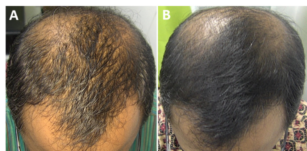
研究背景:雄激素性脱发(AGA)是全球最常见的脱发类型,传统药物(米诺地尔/非那雄胺)或 PRP 对部分人群不适用或依从性差;本研究评估 675-nm 红光激光(RedTouch,
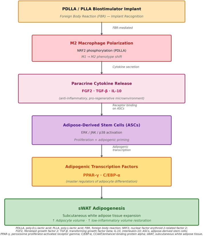
背景:GLP-1 受体激动剂减重常伴随“Ozempic Face”式面部容积流失与皮肤松弛,其力学机制为张力稳态崩溃;透明质酸(HA)填充剂以占位为主、储存模量(G′)偏低,难以持