IncobotulinumtoxinA上面部精细注射策略:超稀释+微剂量+扩展点位兼顾自然表情
2026年8月8日36 min read
Clinical, Cosmetic and Investigational Dermatology 2025;18:608735
Clinical, Cosmetic and Investigational Dermatology 2025;18:608735
The eyebrow is a central anatomical unit in upper facial aesthetics, where multiple muscle groups—including the frontalis, corrugator supercilii, and orbicularis oculi—interact to influence its position and movement. Achieving an optimal balance among these muscles is crucial to enhance facial harmony while preserving natural expressiveness. This study describes an anatomy-based injection strategy using hyperdiluted incobotulinum toxin A, administered through a higher number of injection sites for each muscle in the upper third of the face. Compared with conventional methods using fewer, more concentrated injection points, this approach is intended to promote more uniform toxin distribution, minimise untreated areas, reduce the occurrence of compensatory wrinkles and support balanced neuromodulation while limiting excessive muscle weakening. By integrating microdosing, hyperdilution, and expanded injection mapping, the proposed technique aims to preserve eyebrow dynamics, reduce the risk of brow ptosis, and achieve a more natural aesthetic outcome. The approach is compared with conventional injection patterns, highlighting differences in dose distribution, injection depth, and muscle targeting. Two clinical cases illustrate the feasibility and potential clinical utility of the proposed approach as a refined and customisable strategy for upper facial rejuvenation.
Keywords: aesthetics、eyebrow dynamics、facial harmony、hyperdilution、incobotulinum toxin A、neuromodulation、upper facial rejuvenation
Aesthetic medicine has evolved to meet the growing demand for non-surgical procedures addressing age-related changes. This demand has expanded the use of injectables, particularly minimally invasive neurotoxins and dermal fillers, while advances in formulation, injection technique, and treatment protocols have contributed to improve efficacy and safety. 1
Botulinum toxin type A is a neurotoxic protein produced by Clostridium botulinum that temporarily inhibits acetylcholine release at the neuromuscular junction, resulting in muscle relaxation. 2 Initially approved for the treatment of several neuromuscular disorders, including strabismus, blepharospasm, cervical dystonia, and upper limb spasticity, it has become one of the most widely used treatments in aesthetic medicine. 3 , 4
Despite its success, early aesthetic applications of botulinum toxin were frequently criticised for producing an unnatural or “frozen” appearance characterised by reduced facial expressiveness and loss of facial harmony. 5 , 6 Over time, patient expectations have evolved, with increasing demand for natural-looking results that preserve individual facial characteristics and emotional expression. 7 Natural outcomes have also been associated with improvements in psychosocial well-being and quality of life. 6 , 8 This expectation is also evident among younger toxin-naïve patients, who often express concern about appearing frozen or unnatural following treatment. 9
As cosmetic patient expectations continue to evolve, aesthetic practitioners are increasingly required to achieve meaningful and natural outcomes while maintaining facial balance and function. 8 Achieving this goal requires a detailed understanding of facial functional anatomy, appropriate dosing strategies, and injection techniques tailored to each patient’s individual anatomical characteristics and aesthetic needs. 10 Because facial muscles function as an integrated system, excessive weakening of a single muscle or inadvertent treatment of adjacent structures may disrupt facial harmony and contribute to unwanted aesthetic outcomes. A more refined understanding of muscular interactions therefore allows for greater treatment precision, improved aesthetic outcomes, and potentially fewer adverse effects. 10
The periorbital region represents one of the primary targets of facial rejuvenation procedures, as the eyes and their surrounding structures play a central role in facial attractiveness and interpersonal communication. 11 The appearance of the eyes is influenced not only by the eyelids themselves but also by the forehead, eyebrow position, and glabellar complex, all of which contribute to the overall perception of facial expression and attractiveness.
Among these structures, the eyebrow occupies a key aesthetic and functional position. Its shape and position are determined by the dynamic balance between the frontalis muscle, the sole elevator of the eyebrow, and several depressor muscles, including the corrugator supercilii, procerus, and orbicularis oculi muscles. These muscles converge within the glabellar and periorbital regions and exert coordinated effects on both eyebrow position and facial expression. Their interaction also contributes to the formation of characteristic facial rhytids, including forehead lines, glabellar lines, and lateral canthal lines. Consequently, successful treatment of the upper third requires careful modulation of the balance between eyebrow elevators and depressors rather than isolated treatment of individual muscles. 12
Reconstitution volume and dilution influence the onset, efficacy, and duration of botulinum toxin type A treatment. 6 Small volumes of highly concentrated neurotoxin may limit distribution within the target muscle, whereas higher dilution combined with multiple injection sites may promote a more homogeneous intramuscular distribution. Such an approach can be adapted to individual anatomical characteristics and treatment goals, supporting natural facial balance and expression. 5 , 6 , 13
Incobotulinum toxin A (Bocouture ® , Merz Pharmaceuticals GmbH, Germany) is a formulation free from complexing proteins and containing only the active neurotoxin. 14–17 Its characteristics support precise treatment planning and make it suitable for personalised injection strategies.
Research suggests an optimal dose threshold related to receptor saturation: 18 , 19 higher concentrations allow more neuromuscular junction receptors to bind the neurotoxin and, once saturated, further dose increases do not yield additional benefits. 6 , 20
This article describes an anatomy-based treatment approach developed by the author over 14 years of clinical practice for the management of the upper third of the face using incobotulinum toxin A. The proposed strategy combines hyperdilution, microdosing, and an expanded injection pattern to optimise neuromodulation while preserving facial expression and eyebrow dynamics. The technique aims to enhance facial harmony, minimise the risk of asymmetries and eyelid ptosis, and reduce treatment-related adverse effects. Two clinical cases are presented to illustrate the feasibility and practical application of this approach in routine clinical practice.
The natural equilibrium between agonist and antagonist muscles is disrupted by neuromodulators. Therefore, before treatment, the practitioner must have a comprehensive understanding of function, structure, and localisation of facial muscles.
The face should be assessed with the eyes open and closed, at rest, with moderate animation, and under maximum contraction. Neurotoxin-induced temporary muscle paralysis reduces both dynamic and static wrinkles. 2 , 10
Incobotulinum toxin A is supplied as a powder in vials containing 50 or 100 units (U) of purified incobotulinum toxin A (150 kD). It must be reconstituted with 0.9% sodium chloride solution (9 mg/mL) prior to use, in accordance with the manufacturer’s prescribing information. 21
The reconstitution protocol includes: Cleaning the vial’s rubber stopper with 70% alcohol before needle insertion.Inserting the needle vertically through the rubber stopper to allow vacuum-assisted withdrawal of the saline solution, and gently injecting any remaining saline into the vial, if necessary, to avoid foam formation.Removing the syringe and mixing the solution by gentle swirling and inverting the vial, avoiding vigorous shaking. 21 , 22
Incorrect reconstitution may reduce efficacy and compromise treatment success. 22 For example, foam formation, typically caused by excessive shaking, may indicate protein denaturation, which can reduce the number of active units available.
Prescribing information outlines several reconstitution volumes for 50 U and 100 U vials. Reconstituting a 50 U vial with 1.0 mL of sodium chloride 9.0 mg/mL (0.9%) solution for injection yields a concentration of 5 U per 0.1 mL, whereas reconstitution with 1.25 mL provides 4 U per 0.1 mL. Similarly, reconstituting a 100 U vial with 2.0 mL of solvent yields 5 U per 0.1 mL, and with 2.5 mL results in 4 U per 0.1 mL. 21 In clinical practice, the final dilution is typically based on practitioner preference, generally ranging from 0.25 mL to 5 mL per 100 U. 1 , 7 , 23
In the present technique, the author employs a hyperdilution approach, reconstituting 50 U of incobotulinum toxin A in 1.5 mL and 100 U in 3.0 mL, using a 1.0 mL of solvent and 100-IU insulin syringe without dead space. Each notch corresponds to 3.33 U.
Vial size selection depends on the extent of the treatment area and the number of planned administrations per muscle. Dosing is adjusted according to the thickness of the targeted muscle, which varies between individuals. 24 Injection depth also depends on the objective: deep intramuscular injections promote weakening or transient paralysis of the treated area, whereas superficial injections (ie, above the muscle) target superficial muscle fibres and their connection with the skin. 25
Incobotulinum toxin A is indicated for the temporary aesthetic improvement of upper facial lines, including horizontal forehead lines (at maximum contraction), glabellar frown lines (at maximum frown), and lateral canthal lines (crow’s feet, at maximum smile), in adults under 65 where the severity has a psychological impact. 21
Facial muscles function in coordinated complexes and interact closely with adjacent anatomical components. 26
A recent anatomical study identified eight distinct forehead layers: skin, superficial fatty layer, suprafrontalis fascia, the orbicularis oculi and frontalis muscles, a homogeneous fat layer (including preseptal fat in the upper eyelid, retro-orbicularis fat deep to the orbicularis oculi muscle, and retro-frontalis fat deep to the frontalis muscle), subfrontalis fascia, including preperiosteal fat within the prefrontal space in the lower forehead), and periosteum. 26 These findings highlight region complexity, where muscle fibres, connective tissue, and subcutaneous structures are intricately interwoven. 27
The forehead is largely occupied by the frontalis muscle, which is divided medially by a central aponeurosis. 4 , 26 , 28 The frontalis muscle originates posteriorly from the galea aponeurotica and interdigitates anteriorly with the procerus, corrugator supercilii, and orbicularis oculi muscles. 12 , 29 Notably, it lacks bony insertion and origin, is surrounded by superior and a subfrontal fasciae, 12 , 26 and its fibres may extend laterally beyond the hairline. 28
The frontalis main functions (scalp retraction and eyebrow elevation) define the bidirectional movement of the skin of the forehead. 4 These movements converge at the horizontal line (C-line) located roughly two-thirds above the eyebrow. 27 Frontalis contraction produces horizontal lines on the skin which can be straight or curved, depending on muscle morphology. 28 Repeated contractions produce transverse rhytids, influenced by interindividual structural variations. 30
The primary objectives of forehead treatment are to elevate the eyebrow, reduce horizontal rhytids, and minimise the appearance of lateral transverse compensatory wrinkles (eyebrow commas).
The personal approach prioritises eyebrow elevation, particularly in patients with superior palpebral hypertrophy.
The frontalis muscle plays a critical role in eyebrow positioning. The number and placement of injection points are tailored to individual anatomy, including forehead width and mass; factors such as gender, ethnicity, and previous treatments must also be considered.
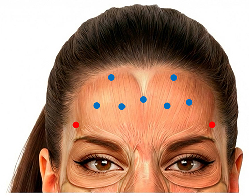
Five injection sites are positioned along the medial inferior portion of the frontalis, approximately 2 cm above the eyebrows. One central point aligns with the C-line, and two lateral sites per side are placed slightly higher, in a V-shape fashion. Each site receives 1.6 U of hyperdiluted product deeply ( Figure 1 ). The number of units and injection sites may vary between patients, and the central site may be omitted where appropriate.
Figure 1 Personal injection technique for the forehead. The frontalis is treated with seven intramuscular injection points and two superficial intradermal points. The central injection point may be omitted, depending on anatomy. Blue circles, Deep injections; Red circles, Superficial injections.额肌:7 个肌内深注射点(蓝)+ 2 个真皮浅注射点(红)。下排 5 点位于眉上约 2 cm,中央点对齐 C 线、可按解剖省略;发际线下另设 2 个外侧深点。每深点 1.6 U,每浅点 0.6 U,总量 12–14 U。Two additional lateral deep injection sites are positioned just below the hairline to treat the upper forehead, which may exert downward force on the eyebrows. Each site receives up to 1.6 U of hyperdiluted product ( Figure 1 ).
To reduce the risk of compensatory wrinkle formation, two intradermal injection sites (one per side) are positioned above the eyebrow tail (1 cm below the hairline), each receiving up to 0.6 U of hyperdiluted product ( Figure 1 ).
In total, 12–14 U of incobotulinum toxin A are administered to the frontalis, lower than conventional techniques (10–20 U), but distributed across more sites and a larger area. This approach is intended to promote a more even distribution of the neurotoxin while limiting the risk of excessive frontalis weakening and eyebrow droop ( Table 1 ).
Table 1 Use of Incobotulinum Toxin A in the Forehead: Conventional vs Personal Technique21
| Conventional Technique | Personal Technique | |
|---|---|---|
| Number of injection sites | 5 (intramuscular) | 9 (7 intramuscular; 2 intradermal) |
| Units per site | 4 U per site (deep injection) | Up to 1.6 U per intramuscular site (7 points)Up to 0.6 U per intradermal site (2 points) |
| Total dose | 10–20 U | 12–14 U depending on frontalis muscle extension and thickness |
The glabellar complex is primarily defined by the interconnection between the procerus and the paired corrugator supercilii muscles. 4 , 31
The procerus runs obliquely from the midline soft tissue over the nasal bone and inserts into the dermis and frontalis muscle near the upper margin of the medial eyebrows. 4 , 31 , 32 Procerus contraction draws the medial eyebrows downward, producing horizontal lines on the forehead. Over time, repeated contractions lead to horizontal wrinkles at the nasal root. 4
The corrugator supercilii originates deeply from the superciliary arch of the frontal bone, approximately 0.5 cm lateral to the midline (eyebrow head). The muscle extends laterally and cranially, inserting into the middle and/or lateral third of the eyebrow skin, where its fibres join those of the frontalis and orbicularis oculi. 31 The corrugator supercilii contraction pulls the eyebrows down, and repeated, prolonged contractions cause vertical glabellar rhytids. 4 , 10 , 30
Together, the procerus and corrugator muscles medialise and draw the eyebrows, opposing the lifting force of the frontalis. 4
The conventional technique typically involves one central injection point into the procerus and two points on each side of the corrugator (medial and lateral), resulting in five intramuscular injections.
Personal technique uses two deep intramuscular injection points per corrugator, the first more medially, and the second more laterally, approximately 0.5 cm above the orbital rim, but not too high, to avoid impacting the frontalis fibres and causing potential brow ptosis ( Figure 2 ).
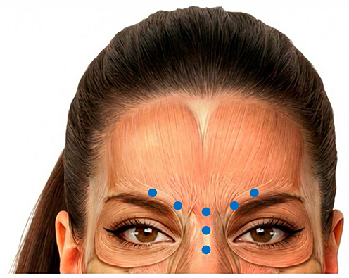
Figure 2 Personal injection technique for the glabellar complex. Deep intramuscular injections: two points per corrugator supercilii (medial and lateral) and three points along the procerus muscle. Blue circles, Deep injections.眉间复合体:每侧皱眉肌 2 点(内侧 3.3 U 深达骨面 / 外侧 1.5 U),降眉间肌沿鼻背垂直排布 3 点、各 1.5 U。常规术式只在降眉间肌打 1 点,会漏掉肌肉下段。The needle is advanced until it reaches the bone and is oriented inferior to superior, and medial to lateral. The medial point receives 3.3 U of hyperdiluted product, while the lateral point is injected with 1.5 U. These sites align anatomically with those in the conventional technique.
Notably, the effect of the injections placed in the inferior frontalis region also extends to the glabellar region.
The procerus is treated using three intramuscular points, rather than a single central point injected with 4 U of non-hyperdiluted product in the standard approach. The points are aligned vertically along the central portion of the nose and extend below the nasal root to facilitate treatment of the entire muscle, including its lower portion, an area missed in the conventional method. Each point receives 1.5 U of hyperdiluted product ( Figure 2 ).
Overall, the total dose for the procerus ranges between 4 and 5 U, while the total dose for both corrugator muscles is approximately 9 to 10 units, depending on individual morphology and muscle mass ( Table 2 ).
Table 2 Use of Incobotulinum Toxin A for the Glabellar Complex: Conventional vs Personal Technique21
| Conventional Technique | Personal Technique | |
|---|---|---|
| Number of injection sites | 5 (intramuscular) | 7 (intramuscular) |
| Units per site | 4–6 U | For each corrugator muscle:- 3.3 U, medial, deep- 1.5 U, lateral, more superficial injectionProcerus muscle:1.5 U at each of the three points |
| Total dose | 20–30 U | 14–15 U |
Although the total units administered to the glabellar complex exceed the conventional technique, hyperdilution is intended to promote a more homogeneous distribution and action across all muscle fibres.
Located in the subdermal layer, the orbicularis oculi surrounds the upper and lower eyelids in a concentric pattern, and can be divided into three parts, orbital, preseptal, and pretarsal. The muscle is anchored to the bone by the orbicularis retaining ligament. 11
The superficial horizontal fibres of the superolateral portion of the orbicularis oculi help to create vertical glabellar lines by amplifying the effect of corrugator supercilii contractions. The orbicularis oculi is responsible for eye closure during contraction. 4 Repeated contractions of the orbital portion may lead to lateral canthal lines (crow’s feet) during smiling and squinting which may become static with ageing and environmental factors. 33 Repeated contractions of the lower pretarsal portion of the orbicularis oculi muscle may lead to lower eyelid hypertrophy.
In the conventional technique, the orbital portion of the orbicularis oculi and associated crow’s feet are treated with three deep intramuscular injections per side. The upper and lower points are placed along the orbital rim, while an intermediate point is placed approximately 1 cm lateral to and outside the bony orbital rim, aligned with the lateral canthal line.
The personal technique aims to modify this approach by relocating the injection points to optimise distribution, enhance safety, and account for anatomical considerations, particularly in patients over 40 years of age.
The lowest injection point is placed directly at the lateral canthal line, with the remaining two points positioned slightly higher, less than 1 cm apart ( Figure 3 ). All three points are aligned along the bony orbital rim, avoiding peripheral placement. This alignment helps keeping the product within the muscle body, as the effectiveness of hyperdiluted product may be reduced when injected beyond the bony orbital margin.
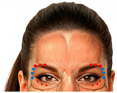
Figure 3 Personal injection technique for crow’s feet and brow lift. Blue circles, Deep injections; Red circles, Superficial injections; Small red circles, Very superficial microinjections.眶周:3 个肌内深点全部沿眶骨缘排列、最低点不低于外眦;眉弓弧线上 3 个真皮内点(各 1.5 U)用于处理眼轮匝肌上外侧份;下睑细纹处 3–4 个极浅微滴(不足 1 U)。The needle is oriented from inside out, nearly parallel to the facial plane, to ensure that the product remains within the orbital rim boundaries.
This higher placement is also based on anatomical considerations. In patients over 40 years of age, the risk of developing lower eyelid bags increases. Injecting too low along the orbital rim has been hypothesised to contribute to this issue in susceptible patients. From an anatomical perspective, excessive weakening of the lower orbicularis oculi may reduce muscular support, potentially leading to detachment from the underlying bone and muscle relaxation. To mitigate this risk, injection points should not be placed below the lateral canthus, preserving the supportive function of the orbicularis while reducing the risk of lower eyelid bags’ development.
The corrugator and superolateral orbicularis oculi play a significant role in frowning. If only the corrugator is weakened, overpowering contraction of the superolateral orbicularis may medialise the eyebrow, pushing the eyebrow head inward. Based on the author’s clinical observations, untreated activity of the superolateral orbicularis oculi may contribute to medial eyebrow displacement and the appearance of vertical compensatory wrinkles in some patients. To address this possibility, the personal technique includes three additional intradermal points in the superolateral portion of the orbicularis oculi along the eyebrow arc, very close to the eyebrow arch, to minimise diffusion into the frontalis and reduce eyebrow drooping without increasing the risk of brow ptosis. Each site receives 1.5 U of hyperdiluted product ( Figure 3 and Table 3 ). The most proximal point, near the temporal crest, may be placed slightly deeper, according to the author’s clinical judgement, with the aim of supporting eyebrow tail elevation and overall aesthetic balance ( Figure 3 ).
Table 3 Use of Incobotulinum Toxin A for Crow’s Feet: Conventional vs Personal Technique21
| Conventional Technique | Personal Technique | |
|---|---|---|
| Number of injection sites (per side) | 3 (intramuscular) | 9–10 total:3 intramuscular;3 intradermal;3–4 very superficial (microdroplets) |
| Units injected per site | 4 U | 3.3 U (hyperdiluted), intramuscular1.5 U intradermalFraction of a unit (microdroplets of hyperdiluted product) |
| Total dose | 12 U per side (24 U total) | ~15 U per side (~30 U overall) |
To treat small lower eyelid wrinkles, the personal technique adds three to four very superficial microinjections, delivered as microdroplets (fractions of a unit), at the origin of each wrinkle ( Figure 3 ). This approach is not included in the conventional technique.
In the conventional approach, each injection site receives 4 U of incobotulinum toxin A, totalling 12 U per side (24 U overall for both orbicularis muscles). In contrast, the personal technique administers 3.3 U of hyperdiluted product at each of the three deep intramuscular points, targeting the orbital portion of the orbicularis oculi, 1.5 U at each of the three superficial intradermal points along the eyebrow arc at the superolateral portion of the orbicularis oculi, and microdroplets at three to four very superficial points at the origin of lower eyelid wrinkles, totalling 15 U per side and 30 U in total ( Table 3 ).
The first follow-up is typically scheduled 20−30 days after the initial treatment session, when the neurophysiological response to botulinum toxin has generally stabilised. In some patients, a touch-up may be beneficial to fine tune results. This may be due to several factors, including potential inaccuracies in dose administration, patient manipulation that may inadvertently displace the product from its intended location, and the need to address small irregularities by adding one or two additional injection points.
This two-stage approach may facilitate greater treatment precision and individualisation, allowing minor adjustments to be performed when clinically indicated.
The proposed technique was developed and progressively refined over 14 years of clinical practice. To illustrate its practical application, two representative clinical cases are presented below. Both patients featured in the images here were treated in accordance with Good Clinical Practice and the ethical guidelines outlined in the Declaration of Helsinki. They also provided informed consent for the publication of their full-face images, as well as the use of their collected data.
Two female patients (aged 34 and 40 years, respectively), with no previous history of aesthetic medical treatments, underwent treatment of the upper facial third using the personal injection approach. Both patients were in good general health, with unremarkable medical histories and no reported allergies or relevant comorbidities.
Post-treatment photographs were obtained at 1 month, with additional follow-up images at 3 and 6 months. Wrinkle severity and eyebrow position were assessed at baseline and during follow-up using the Merz Aesthetic Scales. 34
Both patients were treated using the same standardised injection protocol, targeting the frontalis, corrugator supercilii, procerus, and orbicularis oculi muscles, combined with conservative dosing and adjunctive intradermal microdroplet injections. As the technical aspects of the procedure are detailed in the Materials and Methods section, they are not repeated here.
Static Frontal View (Forehead Lines at Rest)In both patients, frontal wrinkles at rest were markedly reduced up to complete resolution at 1 month. A global elevation of the eyebrows was observed in both patients at follow-up, involving the entire brow rather than only the lateral segment. This finding may reflect the combined treatment of the corrugator supercilii and the superolateral portion of the orbicularis oculi, together with a conservative dosing strategy applied to the frontalis.
At 1 month: Frontal wrinkles improved to grade 0 in both patients (from grade 2 and 3 at baseline, respectively) ( Figures 4 and 5 ).Corrugator wrinkles at rest improved from grade 1 to grade 0 in both cases ( Figures 4 and 5 ).Eyebrow position improved from grade 1 to grade 0 in Patient 1 ( Figure 4 ) and from grade 3 to grade 1 in Patient 2 ( Figure 5 ).
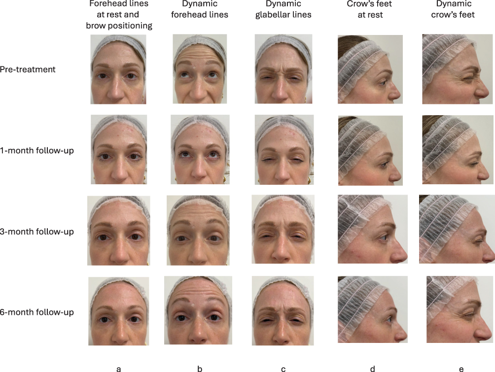
Figure 4 Patient 1: Clinical outcomes following treatment using the proposed injection technique. (a) Static frontal view; (b) Forced frontalis contraction; (c) Forced corrugator contraction; (d) Static lateral view; (e) Forced orbicularis oculi contraction.患者 1(34 岁):静态额纹 2→0 级,眉间纹 1→0 级,眉位 1→0 级;6 个月时额纹轻度复现(1 级),眉位仍维持 0 级。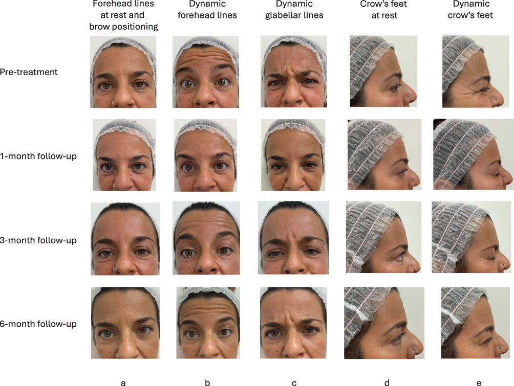
Figure 5 Patient 2: Clinical outcomes following treatment using the proposed injection technique. (a) Static frontal view; (b) Forced frontalis contraction; (c) Forced corrugator contraction; (d) Static lateral view; (e) Forced orbicularis oculi contraction.患者 2(40 岁):静态额纹 3→0 级,眉位 3→1 级;6 个月时额纹回到 2 级、眉位回到基线,衰减快于患者 1。At 3-months, results remained stable in both patients ( Figures 4 and 5 ).
At 6 months, partial attenuation of effect was observed, although outcomes remained superior to baseline in both cases. Patient 1 showed a mild recurrence of frontal lines (grade 1) with eyebrow position maintained at grade 0 ( Figure 4 ). Patient 2 exhibited a more pronounced attenuation of frontal lines (grade 2), with eyebrow position returning to baseline (grade 3) ( Figure 5 ). Corrugator wrinkles remained at grade 0 in both patients ( Figures 4 and 5 ).
Forced Frontalis Contraction (Dynamic Forehead Lines)During forced frontalis contraction, both patients demonstrated complete resolution of dynamic forehead lines at 1 month, with preservation of symmetrical residual brow elevation. These observations were obtained using a relatively conservative dosing strategy, with residual muscular activity remaining visible during facial animation.
On the Merz scale: Patient 1 improved from grade 4 at baseline to grade 0 at 1 month and Patient 2 from grade 4 to grade 1 ( Figures 4 and 5 ).At 3 months, mild recurrence was observed (grade 2 in both cases) ( Figures 4 and 5 ).At 6 months, worsening was recorded in both patients (grade 3) ( Figures 4 and 5 ).
Forced Corrugator Contraction (Dynamic Glabellar Lines)In both patients, the forced corrugator contraction showed complete and harmonious resolution of vertical glabellar wrinkles at 1 month. The simultaneous treatment of the corrugator supercilii and the superolateral orbicularis oculi, both contributing to medial brow traction, may have contributed to limiting compensatory vertical wrinkles related to residual orbicularis activity. At 1 month both patients improved from grade 3 to grade 0 ( Figures 4 and 5 ).At 3 months, a mild reduction in effect was noted in patient 1 only (grade 1) ( Figure 4 ).At 6 months, a minimal recurrence of wrinkles was observed in both patients (grade 2) ( Figures 4 and 5 ).
Static Lateral View (Crow’s Feet at Rest)In the static lateral view, both patients showed eyebrow tail elevation and resolution of crow’s feet at 1 month. Patient 1 improved from grade 1 to grade 0, with results maintained at 3 and 6 months ( Figure 4 ).Patient 2 improved from grade 2 to grade 0, with grade 1 at 3 months and return to baseline severity at 6 months ( Figure 5 ).
Forced Orbicularis Oculi Contraction (Dynamic Crow’s Feet)During forced orbicularis oculi contraction, marked attenuation of crow’s feet was observed in both patients. At 1 month, Patient 1 improved from grade 3 to grade 0 ( Figure 4 ), whereas Patient 2 improved from grade 3 to grade 1 ( Figure 5 ).At 3 months, a slight reduction was noted in Patient 1 (grade 1) ( Figure 4 ), while results remained stable in Patient 2 ( Figure 5 ).At 6 months, both patients showed minimal worsening (grade 2) ( Figures 4 and 5 ).
In frontal views not assessed by the Merz scale, post-treatment images also suggested a reduction in eyebrow head medialisation ( Figure 6 ), a figure that may contribute to the appearance of vertical glabellar lines in some patients. Direct intradermal treatment of lower eyelid wrinkles was associated with visible attenuation of these lines. The anatomical rationale underlying this approach is to minimise weakening of the lower eyelid support structures.
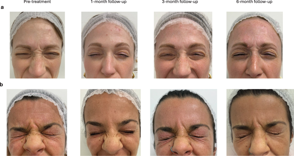
Figure 6 Illustrative reduction in eyebrow head medialisation following treatment using the proposed injection technique. (a) Patient 1; (b) Patient 2.眉头内移(medialisation)减轻。作者认为这正是同时处理眼轮匝肌上外侧份带来的额外收益——只打皱眉肌容易被眼轮匝肌代偿性拉扯眉头。Patient SatisfactionBoth patients expressed satisfaction with the treatment outcome, particularly with the natural appearance achieved and the preservation of facial expressiveness during follow-up.
A key principle of the technique described here is a holistic approach to the muscles of the upper third of the face. These muscles are highly interdependent and work in coordination to create facial expression while maintaining harmony and support. The eyebrow is a key anatomical node where several muscles (frontalis, corrugator and orbicularis) converge and interact; their junctions should be treated appropriately with the aim of promoting eyebrow elevation (primarily via the frontalis) and reducing the risk of downward movement that may result in eyelid ptosis.
While muscle compartmentalisation provides a practical framework, the technique was developed with a recognition of the interconnected nature of facial muscle fibres. To address overlaps and interactions, additional injection points are used beyond standard techniques, with the aim of limiting the risk of asymmetries and other unwanted effects. As Crohn and Greco noted, the frontalis is solely responsible for eyebrow elevation, whereas depression involves the corrugator supercilii, depressor supercilii, procerus, and orbicularis oculi, relationships confirmed through cadaveric studies. 35
The personal technique differs from standard protocols in two respects: 1) a greater number of injection points and 2) the use of hyperdiluted incobotulinum toxin A.
This strategy was designed to promote homogeneous diffusion of the neurotoxin within target muscles, aiming to reduce untreated muscle segments that could contribute to compensatory wrinkle formation. Administering a hyperdiluted solution across multiple injection sites reduces the dose delivered at each injection point and may contribute to a more uniform treatment distribution. 5 This technique may promote a more homogeneous intramuscular distribution of the neurotoxin and has been developed with the aim of optimising treatment efficacy while maintaining a low dose per injection site. As recommended by Cohn and Greco, using the lowest effective dose in the upper third of the face helps prevent undesirable outcomes such as forehead, eyelids, or mouth asymmetry. 35
For example, the frontalis receives 14 U across 10 injection points, compared with up to 20 U across 5 points in the conventional technique. This lower dose may help prevent excessive muscle weakening, thereby minimising the risk of eyebrow drooping.
The personal technique also uses 3 injection points along the procerus (1.5 U of hyperdiluted product per site) rather than a single conventional injection, to favour complete diffusion of the neurotoxin across the full length of the muscle.
Treatment of crow’s feet and the superolateral orbicularis oculi also differs from conventional protocols. Although both approaches use 3 injection points in the orbital portion of the orbicularis oculi, positioning and dosing differ.
In the personal technique, each point receives 3.3 U of hyperdiluted incobotulinum toxin A. The highest injection point is placed near the eyebrow, aligned with the highest dynamic wrinkle and adjacent to the superior bony edge, as close as possible to the orbit; injection sites placed too far laterally may be less effective due to dispersion beyond active muscle fibres.
The lowest point corresponds to the external corner of the eye and is placed centrally on the orbital bone, within the muscle. In the personal technique, this point should not be placed below the lateral canthus, as this may reduce the anchoring function of the lower portion of the orbicularis, causing muscle detachment from the bone, especially in patients over 40 years of age. Such changes have been hypothesised to contribute to the appearance or worsening of infraorbital bags.
Fine lower eyelid wrinkles are smoothed with 3 to 4 very superficial microinjections (microdroplets of hyperdiluted neurotoxin) placed at the origin of each wrinkle. This step, not included in the conventional technique, aims to preserve lower eyelid support.
Injection is guided by anatomy. Deep injections at the origin of the corrugator reach its medial portion and the superolateral orbicularis, whereas more superficial lateral injections address the portion of the muscle that lies closer to the dermis. 35
The personal technique includes a follow-up visit at 20−30 days (approximately 1 month), to allow minor touch-ups for asymmetries, suboptimal dosing or post-treatment manipulation. This first visit is typically followed by two further follow-up assessments at approximately 3 months and about 6 months.
Incobotulinum toxin A was selected because its characteristics support precise treatment planning and individualised injection strategies. 7 , 14 , 17 , 20
This is particularly relevant for younger patients undergoing prejuvenation, a growing demographic seeking natural, preventative aesthetic solutions. 9
The two clinical cases suggest that the proposed injection strategy may provide effective and natural-looking correction of the upper facial third. The similarity of the observations across the two illustrative cases, together with the predictable temporal attenuation of the treatment effect, supports the feasibility of the proposed approach while preserving facial expressiveness.
This proof-of-concept study presents some limitations. Personal approach is one of the existing injection strategies proposed by other authors who deviated from the conventional technique. One example is the refined 3-point injection technique targeting horizontal and vertical glabellar lines described by Cotofana et al. Although differing in execution, both approaches emphasise the integration of anatomic concepts into treatment planning and clinical decision-making. 31
An additional consideration concerns the variability of glabellar contraction patterns and baseline facial asymmetries. Previous authors have highlighted the importance of pattern-based assessment when planning botulinum toxin treatments, as different contraction patterns may require tailored injection strategies. Likewise, asymmetries involving eyebrow position or frontalis activity often necessitate individualised modifications of standard injection schemes. 36 , 37
Although the present article focuses on describing a reproducible anatomy-based injection strategy, it does not specifically address the full spectrum of glabellar contraction patterns or asymmetry-driven treatment adaptations. These factors remain important considerations in clinical practice and may further refine treatment individualisation beyond the scope of the current report. Pattern-based approaches, such as those described by Cotofana and Trindade de Almeida, may provide additional guidance for individual treatment planning and represent a valuable complement to the present technique. 31 , 36 , 37
Furthermore, several authors have so far explored hyperdilution and expanded injection mapping to improve toxin distribution and treatment individualisation. A 6-month prospective, open-label study conducted on 15 women (aged 35 to 65 years) injected with high dilution volume of incobotulinum toxin A in the upper third of face reported improvements in dynamic forehead, glabellar, and lateral periorbital lines over the follow-up period, together with favorable patient-reported assessments. 7
The One21 protocol, implemented by De Sanctis Pecora et al and designed for use with incobotulinum toxin A due to its high precision and low spread, provides an individualised treatment scheme for forehead and glabellar lines based on anatomical references of contraction. The protocol highlights the importance of accurate knowledge of the targeted regional anatomy, precise injection technique, appropriate dose selection and injection depth to improve outcomes and minimise complications. 38–40 Similarly to the present approach, the One21 protocol emphasises individualised treatment planning, detailed anatomical assessment, and the distribution of the neurotoxin according to functional muscle anatomy. 38–40 However, the present technique differs in its use of extensive hyperdilution and in the specific injection mapping developed by the author for the coordinated treatment of the upper facial musculature.
The present article does not propose a novel toxin concept, but rather describes personal anatomy-based implementation of these principles developed over 14 years of clinical practice.
This article describes an anatomy-based injection strategy for the treatment of the upper third of the face using hyperdiluted incobotulinum toxin A distributed across multiple injection points. The two illustrative clinical cases suggest that this approach may support natural-looking outcomes while preserving facial expressiveness. Although limited by the descriptive nature of the report and the small number of cases presented, these observations support the feasibility of the proposed technique and provide a rationale for further investigation in larger patient populations.
先说清楚证据分量。 这是一篇 expert opinion / technique paper, 只有 2 例演示、无对照、无盲法、样本量不足以做任何统计推断。 它的价值不在「证明更好」,而在 把一套注射逻辑讲透了 ——尤其是剂量分布与解剖之间的关系。
三个真正值得琢磨的技术判断:
关于超稀释的争议要如实说: 更高稀释度能否真的带来「更均匀的肌内分布」, 目前仍缺乏高质量的头对头研究支持,主要依据是弥散半径与受体饱和的理论推演。 稀释度越高、扩散范围越大,在眶周这类邻近提上睑肌的区域, 理论上也意味着更高的上睑下垂风险 。 本文用「点位靠骨缘、方向由内向外、真皮内代替肌内」来对冲这个风险,属于经验性的规避策略,而非已验证的安全边界。
可直接用的一条: 治疗后 20–30 天复诊补点,作为常规两阶段流程。 这个时间点神经生理反应已稳定,既能纠正剂量偏差,也能处理患者揉搓导致的产品移位—— 比起一次打满,分两次更容易做出个体化的自然效果。
声明:中文精读 · 仅供学术参考。内容来自公开文献检索,不代表本人观点,不构成诊疗建议。 医疗美容需在正规医疗机构由执业医师实施。
基于相同主题推荐 · 继续深挖