单极射频探头宽度相关组织学差异的比较研究
2026年7月20日14 min read
Lasers in Medical Science · 41:150
Authors: Dong Hye Suh, Sang Doo Lee, Hyun Joo Kim, Kye-Yong Song, Min Kyung Shin, Sang Jun Lee, Hye-Jin Ahn
Journal: Lasers in Medical Science (2026) 41:150 | DOI: 10.1007/s10103-026-04951-z
License: © The Author(s) 2026. This article is licensed under a Creative Commons Attribution-NonCommercial-NoDerivatives 4.0 International License (CC BY-NC-ND 4.0).
Monopolar radiofrequency (RF) devices have improved since 2002, with a significant change in the widths of the tip that comes into direct contact with the skin. We compared the histometric changes between different tip irradiation areas. A total of 28 participants were divided into two groups (14 participants per group) and received a single session of monopolar RF on the face using a 3 × 3 cm² tip (Group S) or 4 × 4 cm² tip (Group L). Skin biopsies were performed before and 2 months after treatment. The densities of collagen and elastic fibers in the papillary, upper reticular, and lower reticular dermis were assessed using the ImageJ software. Group L had a higher collagen density in all dermal layers before and after treatment than Group S; this was particularly noted among those aged 20–39 years. Among those aged > 60 years, Group S had higher collagen density in the papillary dermis than Group L. A large-width tip resulted in deeper and more powerful skin tightening, and a small-width tip may be more suitable for older adults.
Keywords Monopolar radiofrequency · Skin tightening · Collagen fiber · Elastic fiber
Received: 6 April 2026 / Accepted: 5 July 2026
Monopolar radiofrequency (RF) devices use RF energy to heat the dermis and fibrous septae, thus sparing the epidermis from damage [1]. The heat delivered to the dermis causes controlled thermal injury, resulting in rearrangement and regeneration of the dermal matrix [2]. RF heating stimulates fibroblasts to produce new collagen and other proteins that enhance the dermal structure [3]. Monopolar RF treatments improves collagen fiber density with both the CPT (Comfort Pulse Technology) [4] and FLX (Faster, Algorithm, Experience) tips [5]. Advanced since the early 2000s, technological evolution in radiofrequency (RF) systems has progressed from broad energy application to advanced modalities—such as fractional delivery and synergistic multi-modality combinations—enabling precise control over penetration depth and targeted tissue remodeling. Consequently, these continuous methodological refinements have minimized peripheral tissue damage and optimized safety profiles, establishing modern RF technology as a cornerstone of noninvasive anti-aging therapy [6]. Preliminary data and expert opinion suggest that early and repeated intervention with monopolar RF may prevent progressive skin laxity via synergistic and cumulative effects [7]. A significant change in these devices is the difference in tip width, which is in direct contact with the skin. It is regarded that the larger the monopolar RF tip size, the more energy can be delivered to the deeper layers [5]. This study compared histometric quantitative changes according to tip width and different age groups because dermal aging is largely attributed to the accumulation of fragmented collagen and depleted collagen level [7].
The participants were randomly divided into two groups, with each group comprising 14 participants with Fitzpatrick skin types III and IV, aged 24–78 years (mean age, 45 years; 2 men and 12 women per group). Patients with a history of keloid, active cutaneous infections, photosensitivity, uncontrolled general diseases, or insertion of electrical devices such as pacemakers were excluded from this study. Individuals who had undergone energy-based devices, chemical peel, or facial plastic surgery within 6 months were excluded as well. Pregnant or lactating ones were also excluded from this study. Conformed to the tenets of the Declaration of Helsinki, this study (Clinical trial number: not applicable) adhered to the Institutional Review Board (IRB) guidelines, and all participants provided written informed consent. Thermage CPT (Group S) (Thermage® CPT, Thermage® Inc., Hayward, California) and FLX (Group L) (Thermage® FLX, Thermage® Inc., Hayward, California) were used. All participants were informed of possible complications prior to enrollment.
Baseline characteristics including age, Fitzpatrick skin type, and Facial Laxity Rating (FLR) scale score were assessed between group S and L groups [8]. Age was compared using Welch's t-test and Fitzpatrick skin type distribution was compared using the Fisher–Freeman–Halton exact test because of the small sample size. Because the FLR scale is an ordinal variable ranging from 0 to 9, baseline FLR scores were summarized as median and interquartile range (IQR) and compared using the Mann–Whitney U test. Statistical significance was defined as a two-sided p-value < 0.05.
Local anesthesia was induced by applying a topical cream (Lidocaine; Daehan New Pharm, Seoul, South Korea) 1 h before the treatment. After the application of the conductive fluid, each participant received a single session of monopolar RF with static technique on the face using a 3 × 3 cm² CPT tip or a 4 × 4 cm² FLX tip. The FLX tip was 4.0 cm², which was 33% larger than that of the CPT tip. Skin biopsies were performed before and 2 months after treatment and stained with hematoxylin and eosin, Masson's trichrome, and Victoria blue. The treatment levels were 1.5–3.0, and the total delivered energy was 30–45 kJ/cm². 600 shots were administered to the face. For areas with severe erythema, the treatment was resumed once the erythema returned to a normal level or slightly red-color, treatment was stopped in areas where severe erythema persisted, which is presumed as a clinical endpoint. Two dermatologists blindly assessed the dermal density of collagen and elastic fibers in the papillary, upper reticular, and lower reticular dermis using the ImageJ software. The Kruskal–Wallis test was performed to compare the differences in collagen and elastic fiber densities between Groups S and L. For statistical tests, significance was set at p < 0.05. Data were analyzed using the SPSS software (version 23.0; SPSS Inc., Chicago, IL, USA).
There were no significant differences in baseline characteristics between S and L groups. Mean age was 45.3 ± 16.5 years in S group and 44.8 ± 16.9 years in L group (p = 0.938). The distribution of Fitzpatrick skin types was comparable between groups (p = 0.730). Baseline FLR laxity scores were also similar, with median scores of 7.0 (IQR, 5.0–8.0) and 6.5 (IQR, 5.0–7.8) in S and L groups, respectively (p = 0.623) (Table 1).
Collagen and elastic fiber densities in the dermis increased after treatment in both groups (Fig. 1). In Group S, the respective mean densities of the collagen fibers before and after the treatment were as follows: 0.65 and 0.68 for the papillary dermis, 0.57 and 0.59 for the upper reticular dermis, and 0.6 and 0.62 for lower reticular dermis, whereas the corresponding values in Group L were 0.65 and 0.72 (p = 0.0891) for the papillary dermis, 0.54 and 0.62 for the upper reticular dermis (p = 0.005), and 0.58 and 0.65 for the lower reticular dermis (p = 0.0848), respectively. Group L demonstrated higher collagen fiber densities in all dermal layers before and after treatment than Group S, with the difference being particularly significant in the upper reticular dermis. However, the differences in elastic fibers were not significant between the two groups. Participants aged 20–39 years in Group L demonstrated significantly higher collagen densities in all dermal layers before and after treatment than in Group S (Table 2). Similarly, participants in Group L aged 40–59 years had higher collagen densities than those in Group S, although the difference was not significant. However, for participants aged 60–79 years, collagen densities were higher in the papillary dermis in Group S than in Group L, but the difference was not significant (Fig. 2).
Table 1 Baseline demographic characteristics
| Variable | CPT (n=14) | FLX (n=14) | p-value |
|---|---|---|---|
| Age (years), mean ± SD | 45.3 ± 16.5 | 44.8 ± 16.9 | 0.938† |
| FLR laxity scale, median (IQR) | 7.0 (5.0–8.0) | 6.5 (5.0–7.8) | 0.623§ |
| Fitzpatrick skin type, n (%) | 0.730‡ | ||
| I | 0 (0.0) | 0 (0.0) | |
| II | 0 (0.0) | 0 (0.0) | |
| III | 0 (0.0) | 2 (14.3) | |
| IV | 13 (92.9) | 11 (78.6) | |
| V | 1 (7.1) | 1 (7.1) | |
| VI | 0 (0.0) | 0 (0.0) |
Data are presented as mean ± standard deviation (SD), median (interquartile range [IQR]), or number (%). † Welch's t-test; § Mann–Whitney U test; ‡ Fisher–Freeman–Halton exact test
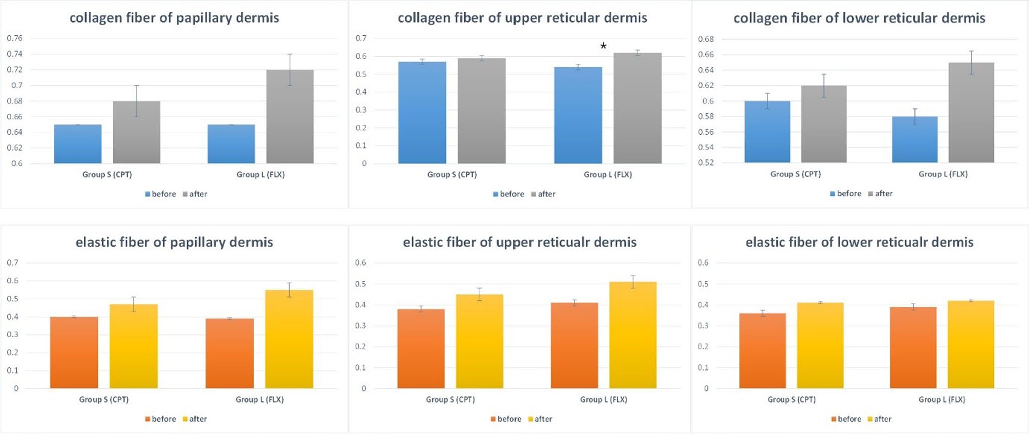
Fig. 1 — Mean densities of collagen and elastic fibers before and after treatment. The densities of collagen and elastic fibers in all dermal layers increase after treatment in both groups. In the upper reticular dermis, the difference in collagen fiber density between before and after treatment differed significantly between Groups L and S (*p < 0.05).
Monopolar RF utilizes electromagnetic waves to generate controlled heat within the dermal layers, stimulating collagen synthesis and remodeling while also promoting tissue contraction [9]. Monopolar RF concentrates electrical energy close to the electrode tip and penetrates to a depth that is about half the electrode's diameter [10]. The FLX tip is larger than the CPT tip, thereby allowing more heat to be delivered faster and deeper into the skin and resulting in heating the dermal tissue to the subcutaneous fat layer. In the monopolar RF treatment, a larger electrode corresponded to a greater treatment depth. Thus, Group L absorbed RF more deeply than Group S and induced more collagen synthesis, which was expected to yield better results. This collagen remodeling is thought to induce a significant collagen tightening effect, thereby manifesting as tangible clinical outcomes, including pore reduction, fine line attenuation, and enhanced skin elasticity and laxity [11].
A previous study on facial skin thickness has reported that the dermal thickness of the forehead, glabella, zygoma, and submandibular skin gradually decreases with age [12, 13]. Because the dermal layer is relatively thinner in older adults, a 3 × 3 cm² tip (Group S), which concentrates energy in a shallower layer, may suit better for delivering energy to the dermal layer. A split-face study on monopolar radiofrequency rejuvenation demonstrated that smaller tips yielded significant improvements in the periorbital, forehead, and glabellar regions. Conversely, larger tips were more effective for treating nasolabial folds and marionette lines [14]. Several recent studies have reported that neocollagenesis begins within days posttreatment, with measurable peaks continuing 2 to 6 months after treatment [7].
Table 2 Differences in collagen fiber density before and after treatment according to age (*p < 0.05)
| Age (years) | Papillary dermis CPT | Papillary dermis FLX | Upper reticular dermis CPT | Upper reticular dermis FLX | Lower reticular dermis CPT | Lower reticular dermis FLX |
|---|---|---|---|---|---|---|
| 20–39 | 0.015 ± 0.09* | 0.134 ± 0.08* | 0.005 ± 0.11* | 0.106 ± 0.07* | 0.001 ± 0.1* | 0.151 ± 0.08* |
| 40–59 | 0.005 ± 0.07 | 0.04 ± 0.14 | 0.005 ± 0.72 | 0.05 ± 0.09 | 0.009 ± 0.08 | 0.021 ± 0.07 |
| 60–79 | 0.058 ± 0.09 | 0.032 ± 0.08 | 0.02 ± 0.15 | 0.096 ± 0.05 | 0.001 ± 0.19 | 0.002 ± 0.1 |
Data are presented as mean ± standard deviation (SD).
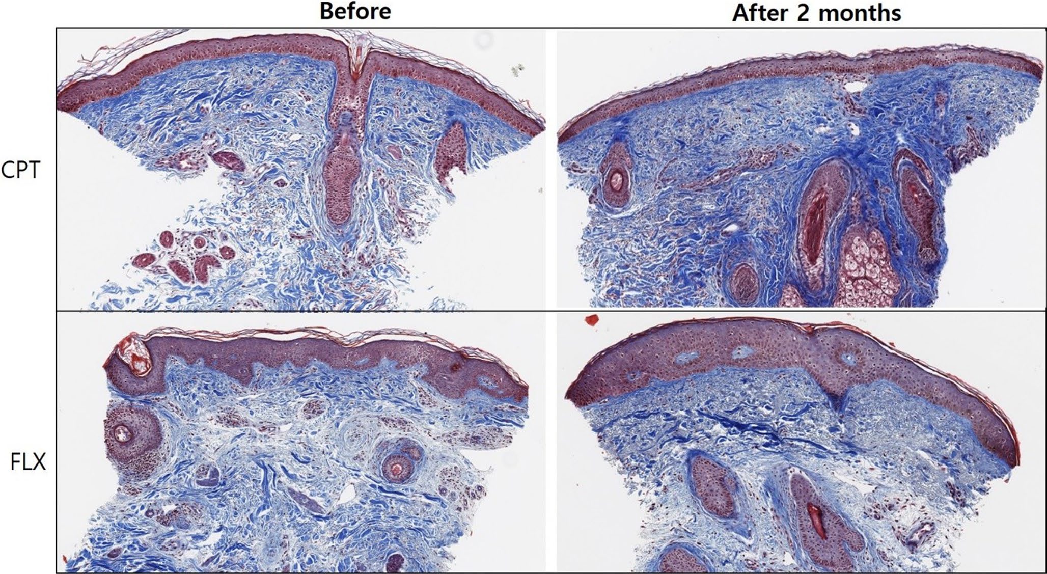
Fig. 2 — Histologic samples derived from older adult participants and stained for collagen fibers using Masson's trichrome; original magnification, ×40. CPT tip-treated sample exhibits a more significant increase in collagen fiber density after treatment.
The limitations of this study included small number of participants and the lack of consideration for clinical changes.
Overall, the 4 × 4 cm² tip (Group L) showed a significant difference in the upper reticular dermis: patients aged 20–39 years had a significantly better large-width effect, those aged 40–59 years had a better but not significantly large-width effect, and those aged 60–79 years had a nonsignificant but better small-width effect in the papillary dermis. In conclusion, the 4 × 4 cm² tip demonstrated a more powerful effect, and the 3 × 3 cm² tip may be a better option for patients aged > 60 years.
Suh D. and Ahn H. conceived and designed the study. Kim H. and Lee S.D. performed the experiments and data collection. Suh D., Ahn H., and Song K. analyzed the data. Shin M. and Lee S.J. contributed materials and analysis tools. Suh D. and Ahn H. wrote the main manuscript text. Kim H. and Lee S.D. prepared the figures and tables. All authors reviewed and approved the final manuscript.
Funding None.
Data availability No datasets were generated or analysed during the current study.
Ethic approval The authors certify that the study was performed in accordance with the ethical standards as laid down in the 1964 Declaration of Helsinki and its later amendments or comparable ethical standards.
Consent to participate/publish All study participants provided informed consent.
Competing interests The authors declare no competing interests.
Open Access This article is licensed under a Creative Commons Attribution-NonCommercial-NoDerivatives 4.0 International License, which permits any non-commercial use, sharing, distribution and reproduction in any medium or format, as long as you give appropriate credit to the original author(s) and the source, provide a link to the Creative Commons licence, and indicate if you modified the licensed material.
📄 下载全文PDF
声明:中文精读 · 仅供学术参考。内容来自公开文献检索,不代表本人观点,不构成诊疗建议。 医疗美容需在正规医疗机构由执业医师实施。
基于相同主题推荐 · 继续深挖
2026/07/20 · 文献笔记 · 14 min read
28名受试者随机分为两组,分别接受3×3 cm²(CPT,Group S)与4×4 cm²(FLX,Group L)单极射频单次治疗,治疗前及治疗后2个月取皮肤活检。结果显示宽探头(4×4 cm²)在各真皮层的胶原密度更高、作用更深更强,尤其20–39岁人群显著;而>60岁人群在乳头层以窄探头(3×3 cm²)更优。结论:宽探头紧致效果更强,窄探头可能更适合老年人。
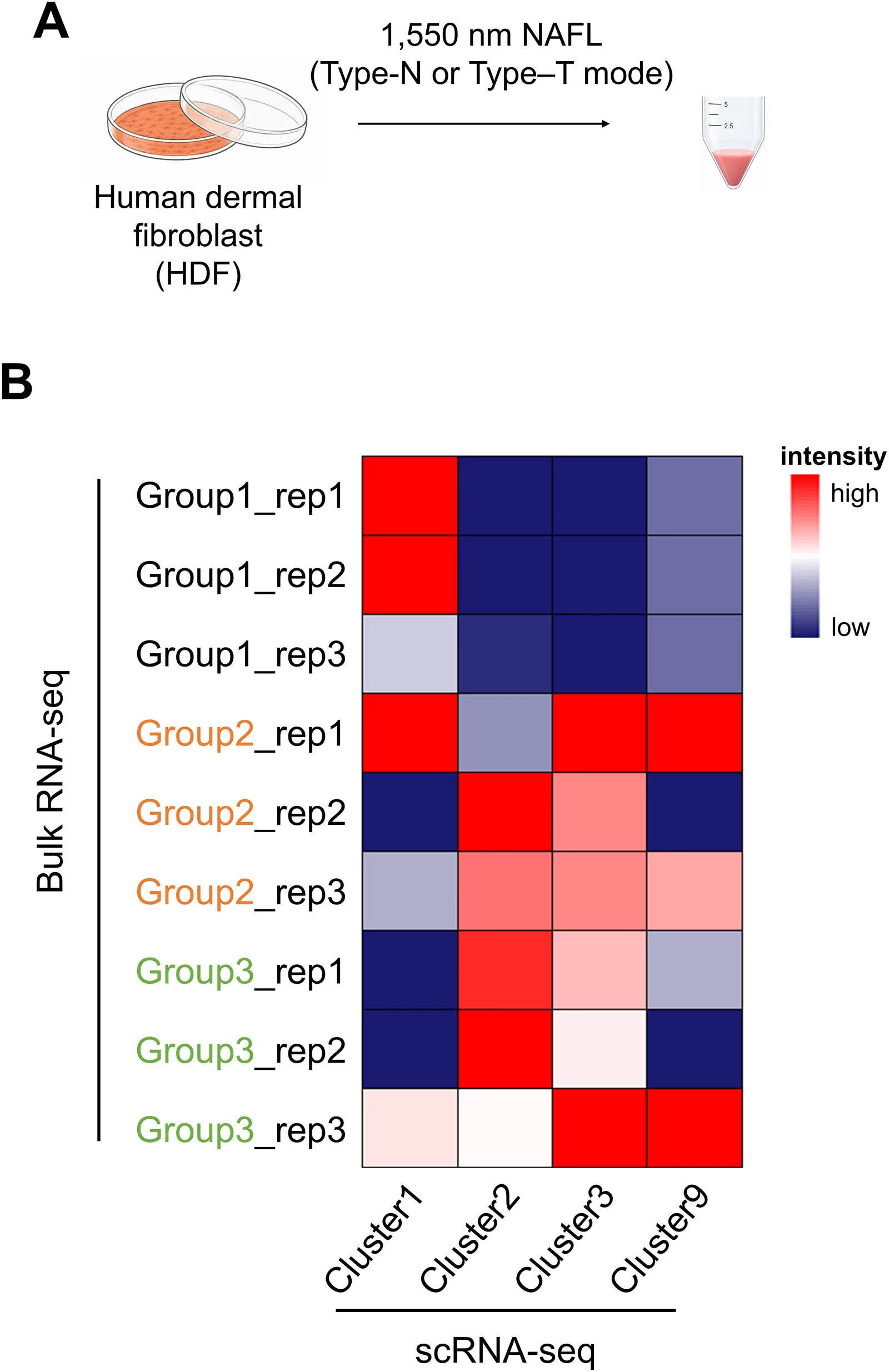
背景:非剥脱点阵激光(NAFL)是常用的真皮重塑与光老化治疗;脉冲时序结构(峰值功率与脉宽组合,总能量密度恒定)对细胞反应的影响研究不足。
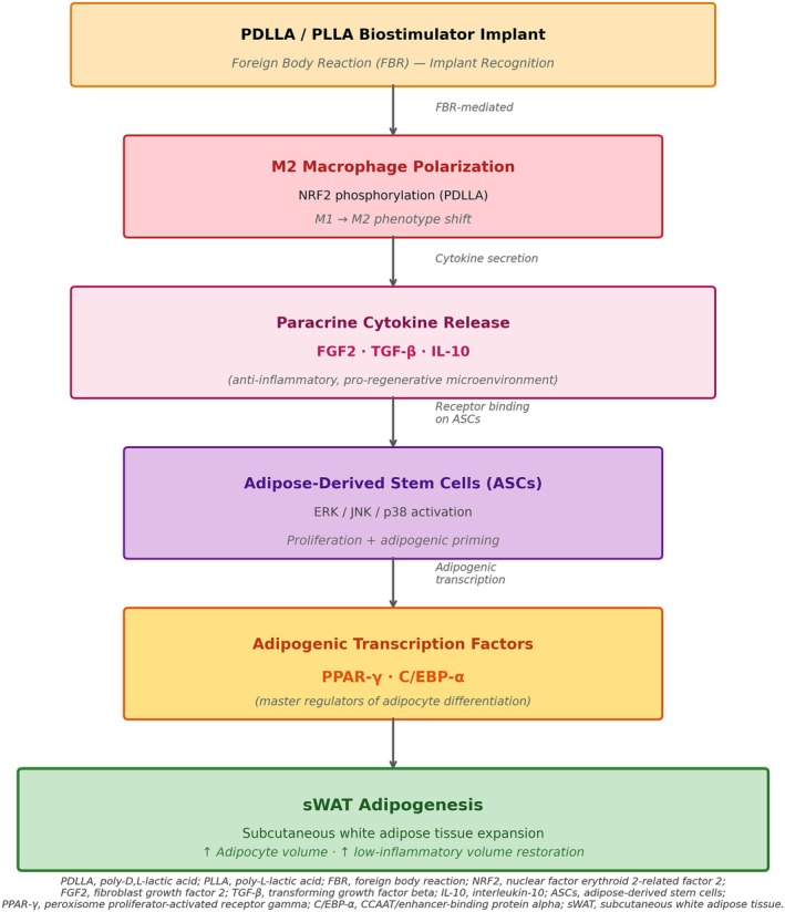
背景:GLP-1 受体激动剂减重常伴随“Ozempic Face”式面部容积流失与皮肤松弛,其力学机制为张力稳态崩溃;透明质酸(HA)填充剂以占位为主、储存模量(G′)偏低,难以持