双路径微聚焦超声面部轮廓与颏下减容
2026年9月9日27 min read
Plastic and Reconstructive Surgery Global Open
Background:Facial aging produces region-specific convexity excess and sulcus depletion, requiring compartmental rather than uniform energy delivery. Dual-mode high-intensity focused ultrasound (HIFU) enables depth- and geometry-selective remodeling of superficial fat and the superficial musculoaponeurotic system.
Methods:In this prospective case series, 38 Korean adults underwent a single session of compartment-programmed dual-mode HIFU across 7 predefined zones (4 facial, 3 submental). Zone-specific intent dictated mode geometry (dot for focal microsupport; linear for broader compaction). Outcomes included 3-dimensional stereophotogrammetry-based volumetry, cervicomental angle, jawline definition index, blinded Global Aesthetic Improvement Scale, and patient satisfaction at baseline, week 4, and week 12.
Results:Thirty-two participants completed week 12 follow-up. Volumetry demonstrated intention-consistent change: central perioral–marionette zone showed microaugmentation (+0.7 cm 3 ), whereas lateral facial zones decreased (zygomatic–nasolabial −2.4 cm 3 ; lateral zygomatic −2.1 cm 3 ; mandibular border −1.5 cm 3 ). Submental volume decreased (central −3.3 cm 3 ; lateral −1.9/−2.0 cm 3 ). Cervicomental angle improved (+6.4°), and jawline definition index increased (+17.1%) ( P < 0.001). At week 12, 93.7% were rated “improved or better” on the Global Aesthetic Improvement Scale; satisfaction was 4.5 ± 0.4. Adverse events were mild and transient.
Conclusions:Compartment-programmed dual-mode HIFU produced coordinated, zone-specific facial and submental contour change by week 12. A longer follow-up is required to confirm durability beyond early remodeling.
Facial contour is governed by the 3-dimensional (3D) distribution of the superficial fat layer and the tension profile of the superficial musculoaponeurotic system (SMAS), both of which undergo region-specific structural changes with age. As the mid- and lower facial adipofascial complex descends, convexities along the zygomatic–nasolabial axis become more pronounced, perioral–marionette depressions deepen, and increasing submental bulk blunts the cervicomental angle, collectively altering facial proportion in a multidirectional manner. These changes reflect not a uniform gain or loss of volume, but a redistribution of mass across regions with distinct biomechanical demands, indicating that effective contour refinement requires layer-directed modulation of superficial fat and SMAS rather than homogeneous lifting.
Accordingly, contour-directed high-intensity focused ultrasound (HIFU) must preferentially target the superficial fat plane (~3.0 mm) and the SMAS complex (~4.5 mm), as these layers exhibit the most predictable shape change in response to focused acoustic energy.
HIFU can deliver focal acoustic energy to predefined depths, generating microthermal coagulation zones within the reticular dermis, fibroseptal network, and superficial adipose layer. 1 , 2 By adjusting focal depth and mode geometry, HIFU can selectively engage the superficial fat plane or the SMAS to induce contraction, neocollagenesis, or adipose remodeling. In contrast to electromagnetic heating technologies—which rely on tissue conductivity—ultrasound-based thermal effects are governed by acoustic focusing and tissue impedance, enabling true layer-selective, depth-specific intervention suited for contour modulation.
Despite these advantages, most prior facial HIFU studies adopted uniform, face-wide parameters, 3 – 6 a strategy that does not reflect the heterogeneity of regional anatomy or the opposing volumetric vectors required for aesthetically coherent mid-to-lower facial improvement. Lateral zygomatic and nasolabial convexities often require targeted fat-plane contraction, whereas the perioral–marionette region may benefit from focal structural support, and submental convexity typically reflects excess in the deep adipofascial compartment. A single homogeneous treatment program cannot satisfy these divergent regional requirements.
Dual-mode HIFU systems address this limitation by modulating irradiation patterns—linear versus dot—which generate distinct thermal geometries. In practical volumetric behavior, the linear mode is intended to induce broader adipofascial compaction consistent with volume reduction (“fat-down”), whereas the low-fluence dot mode is intended to provide focal microsupport and lift-like contour enhancement (“microsupport”), which may present as small volume gains on early 3D imaging during the remodeling window. This directional contrast enables anatomically coherent contour design, aligning the mode with the volumetric requirement of each region. 7 , 8 This provides a mechanistic basis for zone-specific volumetric design rather than generalized tightening.
To translate this principle into a clinically coherent framework, the present study implemented a 7-zone anatomical architecture, consisting of 4 facial zones (zygomatic–nasolabial region, perioral–marionette region, lateral zygomatic region, and mandibular border) and 3 submental zones (central and bilateral). These zones represent the major convexity–concavity determinants of midface, lower face, and submental contour, each characterized by distinct adipofascial thickness, curvature, and volumetric vectors. Such heterogeneity necessitates individualized depth selection and mode allocation rather than uniform, face-wide parameters.
Accordingly, dot mode was used in structurally soft or depleted central zones to provide focal micro-support, whereas linear mode was applied to lateral convex zones to induce broader superficial fat-plane contraction.
Because tissue remodeling after focused ultrasound can continue beyond 12 weeks, we selected weeks 4 and 12 to capture early and intermediate changes while acknowledging that longer-term durability (eg, 6 mo) requires future follow-up. Accordingly, we conducted a prospective study in Korean adults to evaluate whether zone-specific, dual-mode HIFU can induce synchronized and anatomically coherent redistribution of facial and submental volume over a 12-week follow-up period.
This prospective case series was conducted in accordance with the Declaration of Helsinki and was approved by the local institutional review board, with written informed consent obtained from all participants. Thirty-eight Korean adults (30 women, 8 men; age 26–65 y; Fitzpatrick III–IV) seeking nonsurgical facial contour refinement were enrolled (Table 1 ). Eligible facial phenotypes were defined a priori as (1) clinically appreciable lateral midface and/or lower face convexity (eg, zygomatic–nasolabial or lateral cheek fullness) with (2) concomitant contour discontinuity at the mandibular border and/or (3) submental convexity blunting the cervicomental angle. Hollow-prone faces (prominent malar projection with baseline midface concavity, visible skeletalization, or pronounced pre-jowl sulcus at rest) were excluded to reduce the risk of iatrogenic contour irregularity or overreduction. All subjects received a single dual-mode HIFU session. No additional contour-directed energy-based procedures were performed during the study period. Participants were also instructed to avoid neurotoxins, dermal fillers, retinoids (including retinol or tretinoin), collagen-inducing skincare products, and any other aesthetic treatments, including non–energy-based procedures, for 12 weeks following treatment.
Table 1. Baseline Characteristics
| Variable | Value |
|---|---|
| No. enrolled subjects | 38 |
| Gender (female/male) | 30/ 8 |
| Age range (y) | 26–65 |
| Fitzpatrick skin type | III–IV |
| Ethnicity | Asian |
Standardized frontal and lateral digital photographs were obtained at baseline, week 4, and week 12 with a fixed camera distance, identical lighting, and locked International Organization for Standardization/white balance.
Outcomes included 2-dimensional geometric indices—cervicomental angle, which sensitively reflects changes in submental contour and cervicomental definition, and jawline definition index, a measure suited for quantifying mandibular border continuity and lower-facial sharpening.
Three-dimensional volumetric analysis using Vectra stereophotogrammetry was performed to assess zone-specific volume change, enabling direct evaluation of the intended fat-down and fat-up effects across the 7 predefined compartments. Regions of interest were predefined at baseline and duplicated across timepoints, and only minimal pixel correction (<5%) for hair occlusion was permitted.
Two board-certified dermatologists, blinded to the procedure, independently graded the Global Aesthetic Improvement Scale (GAIS), with discrepancies resolved by consensus, whereas patient satisfaction was assessed using a 5-point Likert scale. Statistical significance was defined as a P value less than 0.05.
Pretreatment high-frequency diagnostic ultrasound (L20) was performed to measure dermal, superficial fat, and SMAS depth in each zone, which were used to guide focal-depth selection. Based on these findings, treatment was delivered primarily at 4.5 mm (SMAS level) and 3.0 mm (superficial fat plane), the layers most relevant to contour modulation. Before HIFU delivery, the treatment area was thoroughly cleansed, and ultrasound gel was applied. No anesthesia, cooling, or additional postprocedure care was used.
To reduce the risk of contour irregularity, overreduction, or neuropraxia, treatment was performed using a standardized “safe-plane and safe-stroke” approach: (1) pre-scan each zone with high-frequency ultrasound to confirm target layer thickness and exclude focal hollows, with particular caution in hollow-prone regions (eg, temporal, infrazygomatic, buccal, preauricular areas, and the pre-jowl sulcus) where aggressive compaction may increase the risk of surface irregularity; (2) avoid direct delivery over recognized neurovascular danger zones and bony prominences; (3) maintain consistent transducer contact with adequate gel, perpendicular orientation, and uniform coupling pressure to prevent skip areas and uneven thermal deposition; (4) use nonoverlapping strokes with a spacing approximately equal to one line width, avoiding repeated stacking in thin areas; (5) in convex-to-concave transitions (eg, pre-jowl sulcus), feather the margins by reducing pass count rather than abruptly ending a stroke; and (6) continuously assess skin surface symmetry during treatment and discontinue additional passes if early surface waviness is suspected. Any visible posttreatment contour irregularity was actively screened at follow-up visits and was not observed as a persistent adverse event in this cohort. Zones at increased risk for neurovascular injury that were intentionally avoided during treatment are illustrated in Figure 1 .
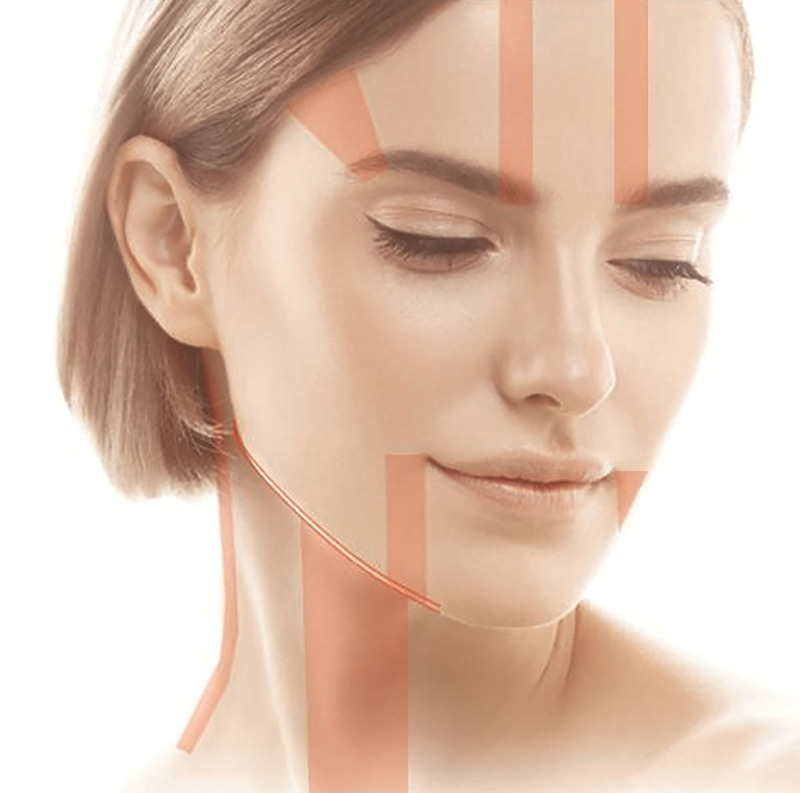
Fig. 1. Major neurovascular pathways and high-risk regions to be avoided during HIFU procedures. Critical anatomical landmarks—including the supraorbital and temporal branches, marginal mandibular nerve, and cervical branch—that require avoidance to minimize risk during energy delivery.HIFU 操作中须规避的主要神经血管通路与高危区域(眶上/颞支、下颌缘支、颈支等)。
A dual-mode HIFU system (LinearZ; Jeisys Medical Inc., Seoul, Republic of Korea) capable of both linear and dot mode was used. Compared with the widely used microfocused HIFU platform (Ulthera/Ultherapy), the present system differs primarily by offering 2 mode geometries (dot and linear) that allow broader compaction strokes versus discrete focal points, enabling compartment-programmed intent (reduction versus microsupport) within a session. Focal depths (3.0 mm superficial fat plane; 4.5 mm SMAS) are conceptually comparable to commonly used ultrasound depths, whereas the present protocol emphasizes zone-specific geometry allocation rather than uniform, whole-face line placement. Patient discomfort was monitored during treatment and recorded as a procedural adverse event category (pain), with most cases resolving within days. Seven anatomical zones were predefined for contour modulation (Fig. 2 ): 4 facial zones (zygomatic–nasolabial, perioral–marionette, lateral zygomatic, mandibular border) and 3 submental zones (central, left, right), each assigned a predefined volumetric intent and mode geometry (dot for focal microsupport; linear for broader compaction).
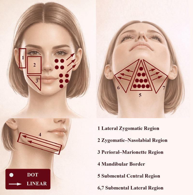
Fig. 2. Seven-zone anatomical treatment map used in this study. Four facial zones and 3 submental zones were predefined, with zone-specific allocation of dot or linear mode according to volumetric intent (microsupport vs reduction). Dot mode (●) represents discrete focal energy delivery for localized microsupport, whereas linear mode (arrows) represents continuous treatment lines applied in a directional manner. Linear strokes were oriented along the natural contour vectors (inside to outside and inferior to superior) to promote tissue compaction and lifting.本研究 7 区解剖治疗图:4 个面部区 + 3 个颏下区,按容积意图分配点状/线状模式。A total of 800 shots were delivered: 400 shots to the facial zones (50 shots per zone × bilateral) and 400 shots to the submental region (200 central; 100 each side), as detailed in Table 2 . Each shot represented a single energy mode event; in dot mode, this corresponded to 1 focal point, whereas in linear mode, it corresponded to a single treatment line.
Table 2. Zone-specific Dual-mode HIFU Programming for Facial and Submental Contour Modulation
| Anatomical Zone | Volumetric Intention | Target Layer and Depth | Mode | Energy (J) | Shot Count | Pass Count |
|---|---|---|---|---|---|---|
| Zygomatic–nasolabial region (mid-face, central) | Reduce convexity (fat-down) | Superficial fat (3.0 mm) + SMAS (4.5 mm) | Dot | 0.4/0.6 J | 50 per side | 1–2 |
| Perioral–marionette region (lower face, central) | Structural support (micro-lift) | Superficial fat (3.0 mm) | Dot | 0.4 J | 50 per side | 1 |
| Lateral zygomatic region | Lateral slimming (fat-down) | Superficial fat (3.0 mm) + SMAS (4.5 mm) | Linear | 0.4/0.6 J | 50 per side | 1–2 |
| Mandibular border | Border sharpening (fat-down) | Superficial fat (3.0 mm) | Linear | 0.4 J | 50 per side | 1 |
| Submental central | Bulk reduction + midline lift | Superficial fat (3.0 mm) + SMAS (4.5 mm) | Dot | 0.4/0.6 J | 200 total | 1–2 |
| Submental lateral (R/L) | Submental tapering (fat-down) | Superficial fat (3.0 mm) | Linear | 0.4 J | 100 per side | 1 |
Mode was assigned according to volumetric requirements: dot mode for central zones requiring focal support and linear mode for lateral zones requiring broader fat-plane contraction, with linear strokes applied directionally from inside to outside and from inferior to superior to follow the natural contour vectors of the jawline and submental region (Fig. 3 ). When 2 passes were performed, additional strokes were delivered in a parallel manner along the same vector, avoiding perpendicular cross-patterning or stacking to prevent excessive energy overlap and uneven thermal deposition. Dot mode strokes were distributed evenly within the target zone without a specific directional orientation.
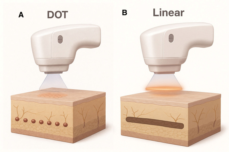
Fig. 3. A, Schematic illustration of dot mode in the dual-mode HIFU system. Dot mode forms multiple discrete micro-coagulation points in the dermal or superficial fat layer, providing localized support and subtle volumization. B, Schematic illustration of linear mode in the dual-mode HIFU system. Linear mode creates an elongated, continuous coagulation line that induces bulk thermal compaction of the superficial fat plane, enabling regional volume reduction and contour refinement.双模式示意:A 点状模式形成离散微凝固点提供局灶支撑与轻微增容;B 线状模式形成连续凝固线诱导体积热压缩。Depth and energy settings were standardized at 0.6 J (4.5 mm; SMAS plane) and 0.4 J (3.0 mm; superficial fat plane). Zones with both superficial fat prominence and SMAS descent (zygomatic–nasolabial and lateral zygomatic) were treated at both depths. The perioral–marionette region received superficial-plane dot strokes only. The central submental zone was treated at both depths, whereas the lateral submental zones were treated at 3.0 mm only (Table 2 ).
Two cartridges were used: 7 MHz for SMAS-adjacent tightening and 4 MHz for superficial fat-plane modulation (Fig. 4 ).
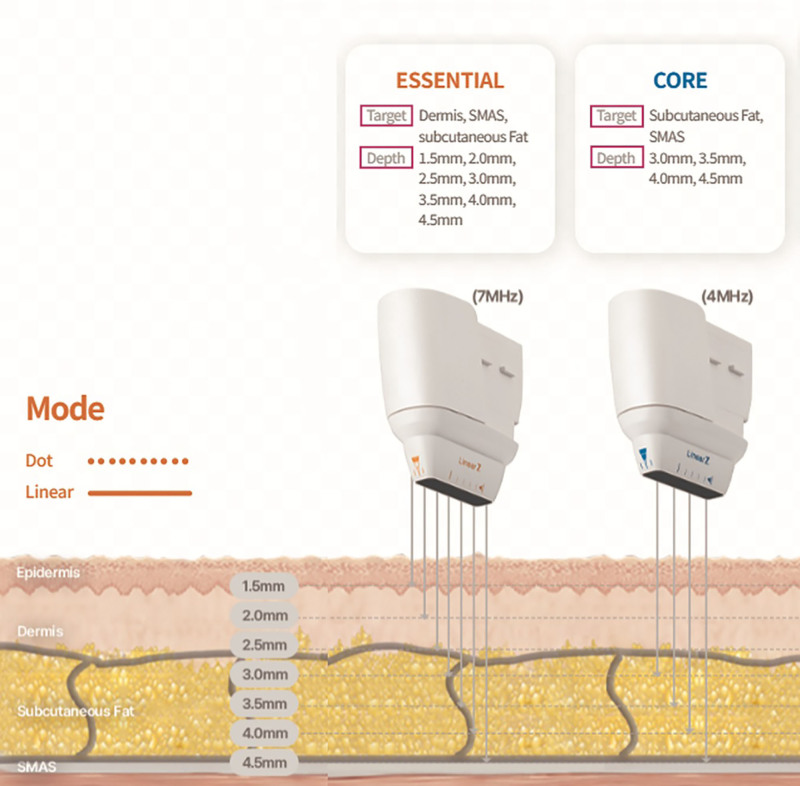
Fig. 4. Depth-specific ultrasound targeting used in the present protocol. The schematic illustrates selectable focal depths corresponding to the dermis, superficial subcutaneous fat, and SMAS layers, enabling compartment-programmed treatment according to anatomical thickness.深度特异性超声靶向:真皮、浅皮下脂肪、SMAS 层的可选焦点深度,实现按解剖厚度的区室化治疗。Treatment was performed from deep to superficial planes with a translation interval approximately equal to 1 line width. Mean procedural time was 15.0 ± 1.5 minutes.
The resulting parameter allocation for each zone—including intention, depth, mode, energy, and shot distribution—is summarized in Table 2 , which outlines the standardized, reproducible programming used for this study.
Thirty-two of 38 participants completed the 12-week follow-up assessment. Directional improvement was already detectable in most subjects by week 4, although early volumetric shifts were modest and variable. By week 12, intersubject variability narrowed and a more consolidated, zone-specific contour response emerged across both facial and submental regions.
Three-dimensional stereophotogrammetry demonstrated intention-consistent mass redistribution.
At week 12, 3D stereophotogrammetry showed zone-specific, intention-consistent volume shifts (Table 3 ): zygomatic–nasolabial −2.4 cm 3 , perioral–marionette +0.7 cm 3 , lateral zygomatic −2.1 cm 3 , mandibular border −1.5 cm 3 , central submental −3.3 cm 3 , and lateral submental −1.9/−2.0 cm 3 .
Table 3. Quantitative Outcomes (n = 32 Completed)
| Outcome Domain | Measure | Week 4 | Week 12 | P |
|---|---|---|---|---|
| 3D volumetry (cm 3 ) | Zygomatic–nasolabial | −0.6 | −2.4 | <0.001 |
| Perioral–marionette | +0.2 | +0.7 | <0.001 | |
| Lateral zygomatic | −0.8 | −2.1 | <0.001 | |
| Mandibular border | −0.6 | −1.5 | <0.001 | |
| Submental central | −1.0 | −3.3 | <0.001 | |
| Submental lateral (R) | −0.5 | −1.9 | <0.001 | |
| Submental lateral (L) | −0.6 | −2.0 | <0.001 | |
| 2D geometry | Cervicomental angle (°) | +1.3 | +6.4 | <0.001 |
| Jawline definition index (%) | +3.2 | +17.1 | <0.001 |
These patterns reflect the planned “central microsupport” (fat-up) and “lateral contraction” (fat-down) logic: central dot-treated zones demonstrated mild volume gain or lift-like behavior, whereas linear-treated lateral zones showed consistent volume reduction and tapering.
Two-dimensional metrics showed parallel shape improvement:
Angle sharpening aligned with the reduction of central and lateral submental volume, whereas the improvement in jawline definition corresponded to mandibular border contraction and smoother mid-lower facial transition.
Representative cases include standardized frontal photographs demonstrating reduced lateral cheek convexity, improved mandibular border continuity, and decreased submental fullness (Figs. 5 , 6 ).
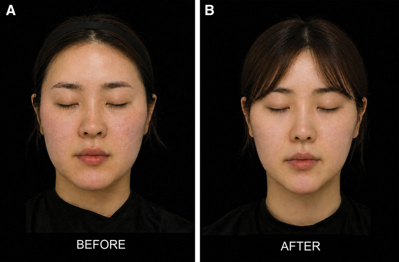
Fig. 5. A, Representative case (woman, 32 years) before treatment. Standardized frontal photograph obtained at baseline before dual-mode HIFU. B, Representative case (woman, 32 y) at 12 weeks after treatment. Standardized frontal photograph obtained 12 weeks after dual-mode HIFU, showing reduced lateral mid-face convexity and improved mandibular border continuity.代表病例(32 岁女性):治疗前与 12 周后面部标准化正位照,示中面部外侧凸度减小、下颌缘连续性改善。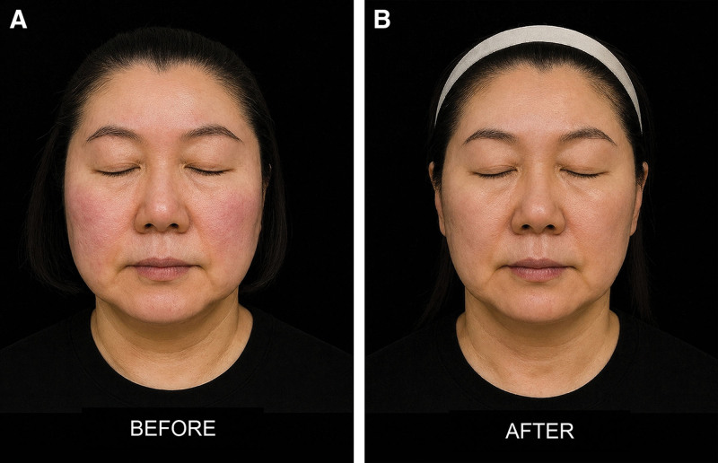
Fig. 6. A, Representative case (woman, 49 y) before treatment. Standardized frontal photograph obtained at baseline before dual-mode HIFU. B, Representative case (woman, 49 y) at 12 weeks after treatment. Standardized frontal photograph obtained 12 weeks after dual-mode HIFU, showing mild but consistent improvement in submental fullness and mandibular contour.代表病例(49 岁女性):治疗前与 12 周后面部照,示颏下饱满与下颌轮廓轻度但一致的改善。
Blinded GAIS ratings indicated “improved” or better in 78.1% at week 4, rising to 93.7% at week 12. Patient satisfaction increased across all zones, with overall scores improving from 4.1 ± 0.6 at week 4 to 4.5 ± 0.4 at week 12, results were often described as: “more definition,” “less heaviness under chin,” “jawline looks clearer.” Zone-specific satisfaction patterns mirrored the treatment design: mandibular border and submental zones showed the greatest gains, whereas central midface regions demonstrated modest but steady improvement.
All 7 zones demonstrated progressive ordinal improvement as shown in Tables 4 and 5 . Improvement trajectories followed the zone-specific intention:
Table 4. Physician Evaluation Scores for the Treated Areas
| Zone | Baseline | Week 4 | Week 12 | P |
|---|---|---|---|---|
| Zygomatic–nasolabial | 2.10 ± 0.58 | 1.92 ± 0.56 | 1.41 ± 0.51 | <0.001 |
| Perioral–marionette | 2.28 ± 0.62 | 1.88 ± 0.57 | 1.33 ± 0.48 | <0.001 |
| Lateral zygomatic | 2.21 ± 0.60 | 1.82 ± 0.55 | 1.29 ± 0.47 | <0.001 |
| Mandibular border | 2.18 ± 0.59 | 1.78 ± 0.53 | 1.25 ± 0.45 | <0.001 |
| Submental central | 2.35 ± 0.67 | 2.05 ± 0.64 | 1.45 ± 0.54 | <0.001 |
| Submental lateral (R/L) | 2.29 ± 0.64 | 1.98 ± 0.62 | 1.43 ± 0.52 | <0.001 |
| Overall | 2.23 ± 0.61 | 1.90 ± 0.57 | 1.36 ± 0.50 | <0.001 |
Table 5. Patient Satisfaction Scores for the Treated Areas
| Zone | Week 4 | Week 12 | P |
|---|---|---|---|
| Zygomatic–nasolabial | 3.8 ± 0.7 | 4.2 ± 0.6 | <0.001 |
| Perioral–marionette | 4.0 ± 0.6 | 4.6 ± 0.5 | <0.001 |
| Lateral zygomatic | 4.1 ± 0.7 | 4.5 ± 0.5 | <0.001 |
| Mandibular border | 4.3 ± 0.6 | 4.7 ± 0.4 | <0.001 |
| Submental central | 4.0 ± 0.7 | 4.7 ± 0.4 | <0.001 |
| Submental lateral (R/L) | 4.2 ± 0.6 | 4.6 ± 0.5 | <0.001 |
| Overall | 4.1 ± 0.6 | 4.5 ± 0.4 | <0.001 |
This concordance across qualitative-quantitative outcomes confirms that the dual-mode, zone-specific HIFU design produced anatomically coherent contour change.
Adverse events were mild and transient (Table 6 ), most commonly pain (18%) and erythema (14%); no serious or delayed neuropathic events were observed.
Table 6. Adverse Events
| AE Category | Incidence, % | Course (Duration) |
|---|---|---|
| Any adverse event | 34 | median 2 d (IQR 1–3) |
| Pain | 18 | median 2 d (IQR 1–3) |
| Transient erythema | 14 | median 1 d (IQR 1–2) |
| Itching | 5 | median 1 d (IQR 1–2) |
| Erythema + pain | 3 | median 2 d (IQR 1–3) |
| Mandibular-distribution numbness | 5 | median 3 d (IQR 2–4) |
| Serious AE | 0 | — |
All adverse events resolved spontaneously within days.
Most facial HIFU studies have applied uniform parameters aimed at generalized tightening or single-vector fat reduction, which may not address simultaneous convexity excess and adjacent sulcus depletion. 9 , 10 Our zone-programmed dual-mode approach was designed to align mode geometry with compartment-specific volumetric needs.
Prior submental-focused trials using multilayer HIFU reported measurable reductions but did not attempt bidirectional (fat-down and fat-up) compartmental modulation within a session. 11 , 12
The present study suggests that a single dual-mode, compartment-programmed HIFU session may induce bidirectional volumetric change at 12 weeks, with reductions in zygomatic–nasolabial and lateral zygomatic regions and measurable augmentation within the perioral–marionette compartment. The decrease in midfacial convexity contributed to narrowing of the mid-lower face and improved mandibular continuity, consistent with gains in the jawline definition index. These outcomes align with the zone-specific allocation of modes, wherein linear mode was applied to excessive convexity and dot mode to structurally soft or deficient regions.
The bidirectional profile observed here is unlikely to reflect uniform tightening. Mechanistically, apparent volume reduction may arise from multiple, nonmutually exclusive pathways: thermal coagulation of fibroseptal networks with subsequent compaction; remodeling of superficial adipose lobules within the 3.0-mm plane; and SMAS-associated tightening that re-drapes overlying soft tissue, reducing projected convexity on 3D surface capture. In the submental region, the combined use of superficial-plane compaction (3.0 mm) and midline deep-plane tightening (4.5 mm) may reduce convexity by both decreasing superficial bulk and improving fascial support, thereby sharpening the cervicomental angle. Because inflammatory edema and early fibrosis can transiently influence surface topography within the first 3 months, durability of these mechanisms must be confirmed with longer follow-up (eg, ≥6 mo). The findings indicate that mode geometry, focal depth, and fluence selection can actively steer adipofascial tissues toward divergent volumetric trajectories within a single session. Corresponding changes in cervicomental angle and jawline definition further support that these modifications were macroscopically appreciable and clinically interpretable.
Historically, global parameter sets aimed at generalized lift or contraction were used, leaving sulcus depletion largely unaddressed. 9 , 13 , 14 The present protocol departs from this convention by applying fat-down (higher-energy linear) to convex compartments and fat-up (low-fluence dot) to deficit zones, positioning dual-mode HIFU as a contour-modulating rather than solely tightening modality.
Temporal response patterns further support a remodeling-driven mechanism. Early changes at week 4 were modest yet directionally aligned, consolidating by week 12, consistent with the kinetics of collagen reorganization and adipofascial remodeling. 12 , 15 The observation that opposite compartments moved in opposite directions within the same individuals reduces the likelihood of systemic confounding such as weight fluctuation.
Depth-specific programming and frequency pairing emerged as principal determinants of volumetric directionality. In this study, the 7-MHz cartridge exerted greater influence on SMAS-adjacent connective-tissue remodeling, whereas the 4-MHz cartridge more effectively modulated the superficial fat plane—findings that align with biophysical principles of frequency-dependent thermal localization. 2 , 16 Pretreatment ultrasonography (L20) provided millimeter-scale characterization of dermal and adipose thickness, enabling selection of the appropriate cartridge for both thin- and thick-skinned patients. 17
Parallel improvement across physician panel ratings and patient satisfaction suggests that the volumetric shifts were clinically meaningful. Integrating quantitative and qualitative outcomes, the most pronounced visible improvements were consistently observed in (1) the lateral zygomatic region, reflected in reduced midfacial width, and (2) the submental complex, corresponding to improved cervicomental angle and patient-reported reduction in “lower-face heaviness.”
This protocol appears particularly suited to phenotypes characterized by mid-to-lower facial adiposity combined with sulcus depletion, a presentation frequently observed in East Asian populations. 12 , 18 Dot-mediated microsupport in deficit zones may reduce early dependence on injectable fillers in such morphotypes. However, because this study intentionally excluded hollow-prone regions to avoid iatrogenic volume loss, the protocol requires adaptation for Western facial phenotypes, where pronounced malar projection and zygomatic contour are often aesthetic ideals. Future studies should evaluate whether zone-specific programming can be recalibrated to accommodate these differing cultural and anatomical standards.
Limitations include the single-arm design without sham control, lack of systematic weight monitoring, and follow-up limited to 12 weeks, which captures early-to-intermediate remodeling rather than long-term stability. Because edema resolution, fibrosis maturation, adipocyte remodeling, and fascial reorganization may continue beyond 3 months, longer follow-up (eg, 6–12 mo) is required to confirm whether early contour changes persist, progress, or partially regress. Future controlled studies should incorporate weight-stability criteria, standardized pain scoring, and longer longitudinal imaging, ideally including ultrasound-based thickness measurements alongside 3D surface volumetry.
At 12 weeks, compartment-programmed dual-mode HIFU (LinearZ) was associated with modest but measurable, zone-specific facial and submental contour changes and high patient-reported satisfaction. Longer-term controlled studies are needed to confirm durability and mechanism beyond early remodeling.
The authors have no financial interest to declare in relation to the content of this article.
Informed consent was obtained from all participants, with full disclosure of the study’s purpose, risks, and confidentiality.
This study was conducted in accordance with the ethical principles of the Declaration of Helsinki. Informed consent was obtained from all participants. No identifying patient information was collected or used.
This study was conducted in accordance with the Declaration of Helsinki under prospective data collection with informed consent. All imaging and clinical data were handled in a de-identified manner to ensure patient confidentiality.
循证与证据等级。 本研究为前瞻性单臂病例系列(38 入组、32 完成 12 周随访),证据等级有限;主要结局采用 3D 立体摄影体积测量与盲法 GAIS,颈颏角与下颌线定义指数改善均具统计学显著(P<0.001),但缺乏假手术对照与体重稳定监测,区域化容积变化属“意向一致”而非因果确证。
局限。 随访仅 12 周,仅覆盖早期至中期重塑,炎症性水肿与早期纤维化可在 3 个月内短暂影响表面形态,持久性(≥6 月)待验证;为安全起见主动排除凹陷易发区,故结论主要适用东亚表型,西方面型需重新校准;不良事件轻且自限,但仍须规避神经血管高危区。
可落地提示。 对以中下面部脂肪堆积合并沟槽空虚、颏下饱满为主诉者,可将双模式(点状/线状)分区 HIFU 作为非手术轮廓调节的评估选项;操作中建议以高频超声预扫描定位层厚、采用“安全平面与安全走行”、规避神经血管危险区,并以 12 周为早期疗效观察窗、以更长随访评估持久性。
声明:中文精读 · 仅供学术参考。内容来自公开文献检索,不代表本人观点,不构成诊疗建议。 医疗美容需在正规医疗机构由执业医师实施。
基于相同主题推荐 · 继续深挖
背景:面部老化呈区域特异性凸度增加与沟槽空虚,需分层(浅脂肪层约 3.0 mm、SMAS 约 4.5 mm)而非均一能量;多数既往 HIFU 研究采用全脸均一参数,难兼顾相反容积向
背景:GLP-1 受体激动剂减重常伴随“Ozempic Face”式面部容积流失与皮肤松弛,其力学机制为张力稳态崩溃;透明质酸(HA)填充剂以占位为主、储存模量(G′)偏低,难以持
背景 / 目的:中面部增量后 noticed 的稳定内侧泪沟轮廓,可能被误判为治疗所致;本单病例观察绘制其跨层连续走行,评估眼轮匝肌浅层(OOc)相关成分是否修饰其显影。