下睑不对称初级下睑整形外科算法
2026年9月1日25 min read
Plastic and Reconstructive Surgery Global Open
Background:Lower eyelid asymmetry is common yet often overlooked. This study proposed a morphologic classification and surgical algorithm based on palpebro-malar groove width, anterior-posterior displacement of the inferior orbital rim, and lower eyelid vertical length. It evaluates outcomes based on this classification and algorithm, using sub–orbicularis oculi fat (SOOF) advancement and orbicularis oculi muscle suspension.
Methods:From June 2023 to May 2025, 267 patients who underwent primary lower blepharoplasty were prospectively evaluated. Patients were categorized into 3 major types (5 subtypes) according to palpebro-malar groove width, posterior displacement of the inferior orbital rim, and lower eyelid vertical length. Surgical techniques were customized according to classification type. Postoperative outcomes were assessed using standardized clinical photography, independent evaluator review, and patient-reported satisfaction.
Results:Among 267 patients (mean age, 56.8 y; 40 men, 227 women), 127 (48%) were type I (mild), 134 (50%) type II (severe), and 6 (2%) type III (right-side dominant). The left inferior orbital rim was positioned more posteriorly than the right in 96% (259 of 267). The left palpebro-malar groove was wider in 38%, whereas the right was wider in only 2%. Postoperatively, 251 patients (94%) reported satisfactory correction; 16 (6%) were partially satisfied, mainly due to inadequate lateral SOOF advancement. Independent evaluator assessment confirmed symmetry in 251 cases.
Conclusions:Lower eyelid asymmetry reflects underlying midfacial skeletal discrepancies between the maxilla and zygoma. The proposed classification facilitates preoperative assessment and surgical planning. Algorithm-based SOOF advancement and orbicularis oculi muscle suspension were associated with improvement in lower eyelid symmetry in most patients.
Attaining optimal aesthetic outcomes in lower blepharoplasty requires not only technical precision but also recognition and correction of preexisting asymmetry. Facial asymmetry is ubiquitous among individuals, but lower eyelid asymmetry in particular is often overlooked during preoperative evaluation and surgical planning. Most patients present with concerns related to aging—such as fat protrusion, skin laxity, or tear trough deformity—rather than asymmetry. 1 – 5 Consequently, they may become aware of their preexisting imbalance only after surgery, when residual differences become more apparent.
Although facial and upper eyelid asymmetries have been discussed in the plastic surgery literature, the lower eyelid has remained comparatively underexplored and insufficiently characterized. 6 , 7 Facial asymmetry may be congenital, developmental, or acquired through trauma or disease. 8 , 9 Previous discussions have largely focused on postoperative complications causing asymmetry—such as ectropion, scleral show, or canthal malposition—rather than inherent skeletal or soft-tissue differences. Unrecognized preexisting asymmetry, however, can contribute to patient dissatisfaction and suboptimal surgical outcomes following lower blepharoplasty.
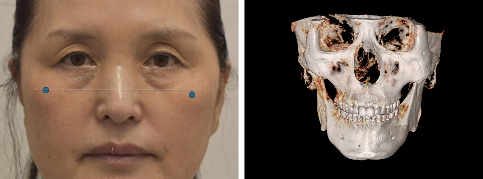
Age-related descent of the lower eyelid and midfacial structures often manifests asymmetrically, producing variability in the tear trough, palpebro-malar groove, and Indian groove. Differences in the underlying osseous framework—particularly the maxilla and zygomatic complex—contribute to these discrepancies in lower eyelid contour and degree of sagging (Fig. 1 ). Prior anatomic studies have reported inconsistent findings regarding laterality—specifically, whether the right or the left side of the face and lower eyelid is wider. 10 – 14 Three-dimensional computed tomography morphometry also confirms zygomatic asymmetry. 15 – 17 According to the AO Surgery Reference diagram, the left orbit lies wider and lower relative to the right. When comparing zygoma prominence, the left side is positioned more laterally in relation to the lateral canthus and positioned lower compared to the right. 18 These skeletal variations result in differential lower eyelid support and variable soft-tissue descent. Therefore, applying the same surgical approach to both lower eyelids will not address the underlying asymmetry, despite an otherwise technically successful lower blepharoplasty (Fig. 2 ).
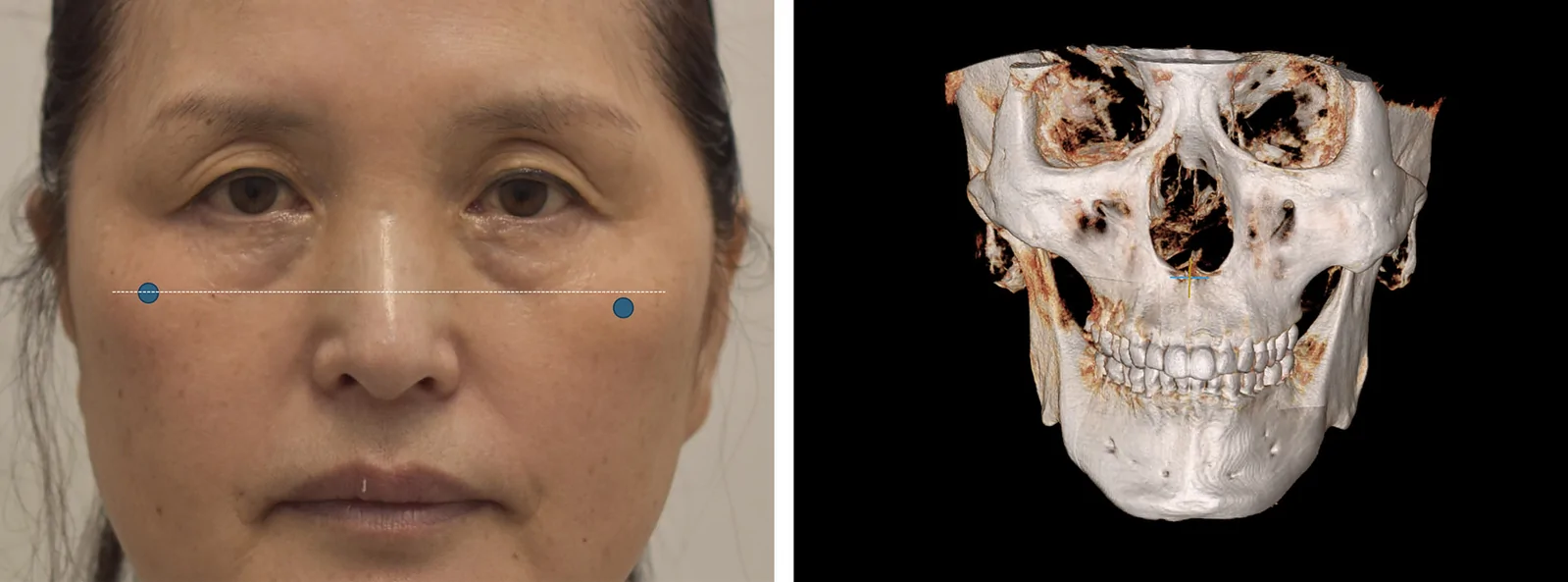
Fig. 1. Clinical photograph and three-dimensional CT reconstruction of facial skeletal asymmetry of the zygomatic complex and maxilla. A, Type I-a classification, 61-year-old patient. Asymmetric facial skeletal structure illustrates the differences in the maxillary and zygomatic contours. Blue dots mark the zygomatic prominence on each side, with the left inferior orbital rim positioned more posteriorly relative to the right. B, Three-dimensional CT reconstruction demonstrating skeletal asymmetry of the zygomatic complex. The left zygoma is wider and positioned more inferiorly relative to the right side.临床照片 + 三维 CT 重建示颧骨复合体与上颌骨不对称(I-a 型,61 岁)。蓝点标记双侧颧突,左下颌缘更后移——直观呈现下睑不对称的骨性形态学基础。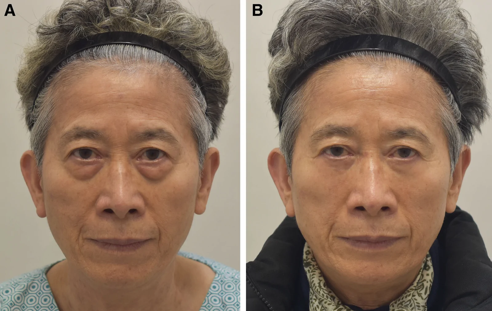
Fig. 2. Type I-b classification, 75-year-old patient, preoperative (A) and 4 months postoperatively (B). Postoperative persistence of lower eyelid asymmetry occurs when preexisting anatomic differences are unrecognized and unaddressed. The left palpebro-malar groove is wider, the inferior orbital rim is positioned posteriorly, and the lower eyelid vertical length is greater, consistent with Type I-b classification. Identical operative techniques were performed bilaterally without adjustment for preoperative asymmetry, resulting in residual postoperative differences. The remaining asymmetry reflects the need for additional SOOF elevation on the left side.I-b 型(75 岁)术前(A)与术后 4 月(B)。示若术前未识别并矫正固有解剖差异,术后下睑不对称可持续存在,强调术前形态学评估的价值。The objective of this study was to propose a morphologic classification system for lower eyelid asymmetry based on palpebro-malar groove width, anterior-posterior displacement of the inferior orbital rim, and vertical length of the lower eyelid. We also describe an algorithmic surgical approach informed by this classification for use in primary lower blepharoplasty.
This prospective study was conducted at a surgery center between June 2023 and May 2025. Consecutive patients undergoing primary lower blepharoplasty with a minimum follow-up of 4 months were included. Patients undergoing revision lower eyelid surgery were excluded. Some patients underwent concurrent procedures, including upper blepharoplasty or forehead lift.
A total of 267 patients were included. Patient age ranged from 32 to 78 years (mean, 56.8 y). There were 40 male and 227 female patients. Follow-up duration ranged from 4 to 24 months (mean, 8.7 mo).
Asymmetry was categorized according to the differences in the width of the palpebro-malar groove, posterior displacement of the inferior orbital rim, and vertical length of the lower eyelid. ( See figure, Supplemental Digital Content 1 , which displays the classification of lower eyelid asymmetry based on differences in the palpebro-malar groove width, posterior displacement of the inferior orbital rim, and the vertical length of the lower eyelid. Each subtype is illustrated with representative images. Shaded areas represent the posterior position of the inferior orbital rim and lower eyelid; darker shading corresponds to greater posterior displacement, https://links.lww.com/PRSGO/F92.)
The lower blepharoplasty skin incision is made bilaterally at the subciliary margin. The skin is elevated off the orbicularis oculi muscle (OOM). The OOM is cut at the junction of tarsal and septal OOM. Through the OOM muscle incision, the dissection proceeds caudally between the suborbicularis muscle fascia and the orbital septum toward the arcus marginalis. In cases where sub–orbicularis oculi fat (SOOF) will be utilized, the dissection continues inferiorly toward the midface by elevating the SOOF in a preperiosteal plane (deep plane undermining) (Fig. 3 ). The herniated orbital fat is transposed inferiorly on both sides with 5-0 Vicryl sutures.
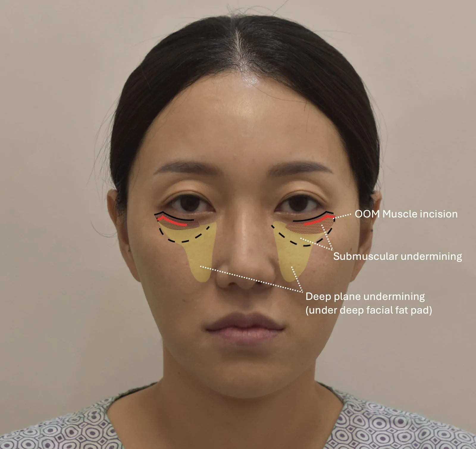
Fig. 3. Schematic illustration showing the surgical approach for SOOF mobilization. Dissection extends inferiorly towards the midface by elevating the SOOF in a preperiosteal plane. The shaded areas indicate the extent of undermining.SOOF 游离手术入路示意图。于 OOM 切口向尾侧在骨膜前平面提升 SOOF 至中面部,阴影示剥离范围,是理解深层平面松解的关键。This technique of incision and dissection applies to all of our patients in our series.We describe the recommended lower blepharoplasty approach based on the subtype of asymmetry (Table 1 ). In general, procedures involved a medial SOOF advancement combined with OOM suspension fixation. Additional modifications, such as an additional lateral SOOF or OOM suspension, were applied based on the severity of asymmetry (Fig. 4 ).
Table 1. Surgical Algorithm According to Asymmetry Type
| Type | Right-side Procedure | Left-side Procedure |
|---|---|---|
| I-a | Medial SOOF advancement + OOM suspension | Same as right side + additional OOM suspension |
| I-b | Same as Type I-a | Same as right side + lateral SOOF advancement + additional OOM suspension |
| II-a | Medial + lateral SOOF advancements + OOM suspension | Same as right side + additional OOM suspension |
| II-b | Same as Type II-a | Same as right side + additional lateral SOOF advancement + additional OOM suspension |
| III | Same as left side + 2 lateral SOOF advancements + additional OOM suspension | Medial SOOF advancement + OOM suspension |
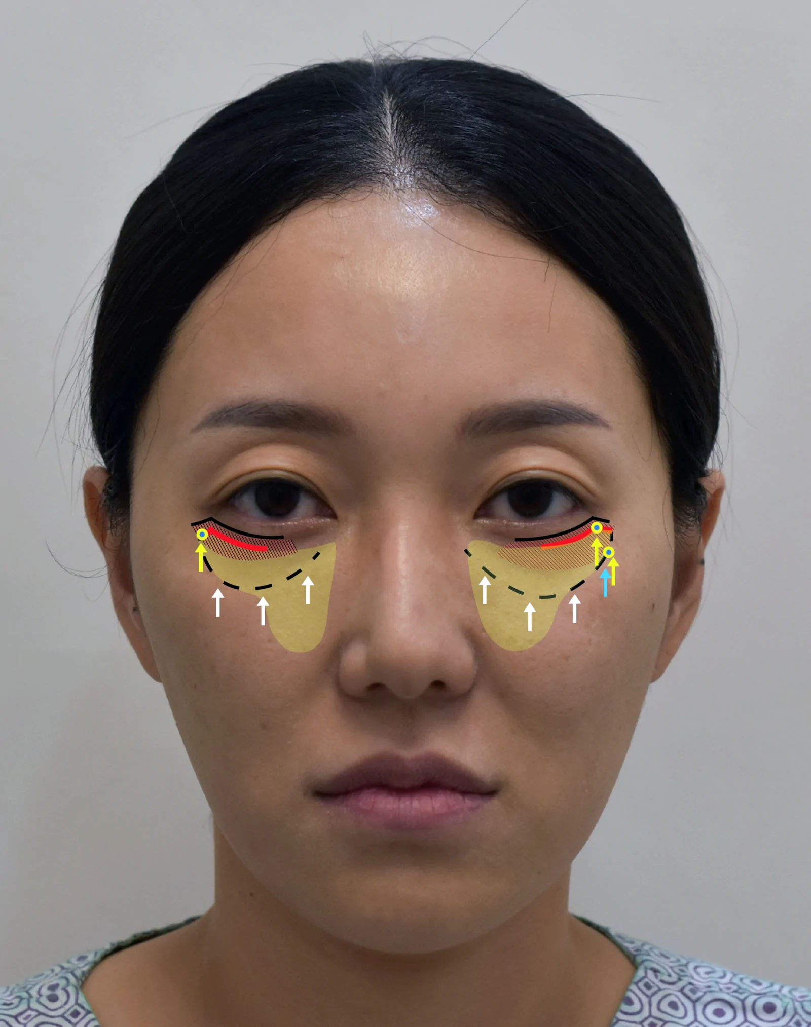
Fig. 4. Schematic illustration of the SOOF elevation and OOM suspension. The medial SOOF is elevated at 2 fixation points, whereas the lateral SOOF may be elevated once or with an additional fixation, depending on the asymmetry type. The SOOF is elevated from medial to lateral at 4 points: (1) deep nasolabial fat pad, (2) medial fat pad, (3) lateral SOOF, and (4) additional SOOF, if required. The white arrows indicate the medial and lateral SOOF. The blue arrow indicates the additional lateral SOOF (in Type II-b cases). The yellow arrows indicate the OOM suspension sites.SOOF 提升 + OOM 悬吊示意图。内侧 SOOF 2 固定点,外侧按需 1 或附加固定;SOOF 自内向外 4 点(1 深鼻唇脂垫、2 内侧脂垫、3 外侧 SOOF、4 附加 SOOF);白箭头示悬吊矢量。算法核心图。For the SOOF elevation, the SOOF is purchased with 5-0 nylon sutures and fixated to the arcus marginalis. The medial SOOF is lifted with 2 fixation points—one in the deep nasolabial fat pad and the other in the medial fat pad. For the narrower palpebro-malar groove side, the medial SOOF is elevated vertically and fixated to the medial arcus marginalis. On the side with the wider palpebro-malar groove, depending on the width, a medial and a lateral SOOF segment or a medial and 2 lateral SOOF segments are elevated and fixated to the arcus marginalis.
For the side with a shorter eyelid vertical length, the OOM is purchased caudally at the level of the lateral orbital rim and elevated cephalically to the inner orbital rim, where it is secured with 5-0 nylon at the horizontal level of the pupil. This upward vector of suspension helps to efface the lateral palpebro-malar groove.
For the side with a greater eyelid vertical length, the OOM is similarly purchased at the level of the lateral orbital rim and elevated cephalically to the inner orbital rim, at the horizontal level of the pupil. As this side requires more elevation, an additional OOM suspension is placed. The additional OOM is purchased just lateral to the initial OOM, at the body of the lateral orbital rim. This dual fixation distributes tension and prevents excessive strain on a single suspension, reducing the risk of a cheese-wiring effect.
To minimize selection bias, all consecutive patients meeting the inclusion criteria during the study period were included. No patients were excluded based on the degree or pattern of lower eyelid asymmetry. Surgical planning was guided by predefined morphologic criteria, including palpebro-malar groove width, anterior-posterior displacement of the inferior orbital rim, and lower eyelid vertical length.
Outcome assessment was performed using standardized preoperative and postoperative clinical photographs obtained during routine follow-up visits. Patient-reported assessment of lower eyelid symmetry was recorded during postoperative evaluations. In addition, postoperative symmetry was independently assessed by two evaluators who were not involved in the surgical procedures. Assessment focused on the presence or absence of residual lower eyelid asymmetry.
Based on our morphologic classification of lower eyelid asymmetry, which incorporates the palpebro-malar groove width, posterior displacement of the inferior orbital rim, and lower eyelid vertical length, and the corresponding surgical algorithm, the results are as follows:
Among the 267 patients evaluated, 127 (48%) were classified as type I asymmetry, including 78 type I-a and 49 type I-b cases. A total of 134 patients (50%) were classified as type II asymmetry, including 80 type II-a and 54 type II-b cases. Six patients (2%) were classified as Type III asymmetry.
In 96% of patients examined, the left inferior orbital rim was positioned more posteriorly than the right. This typically corresponded with an anteriorly rotated right midface and a posteriorly displaced left midface, suggesting an intrinsic skeletal asymmetry favoring posterior displacement of the left side. The left palpebro-malar groove was wider in 38% of cases, whereas the right was wider in only 2%.
Postoperative correction of asymmetry was observed in 251 patients (94%) and they reported satisfactory results (Fig. 5 ). Sixteen patients (6%) reported partial satisfaction, in cases of type II-b or type III, due to incomplete or inadequate lateral SOOF advancement. Independent assessment by two evaluators confirmed the presence or absence of corrected lower eyelid asymmetry, with 251 cases achieving symmetry.
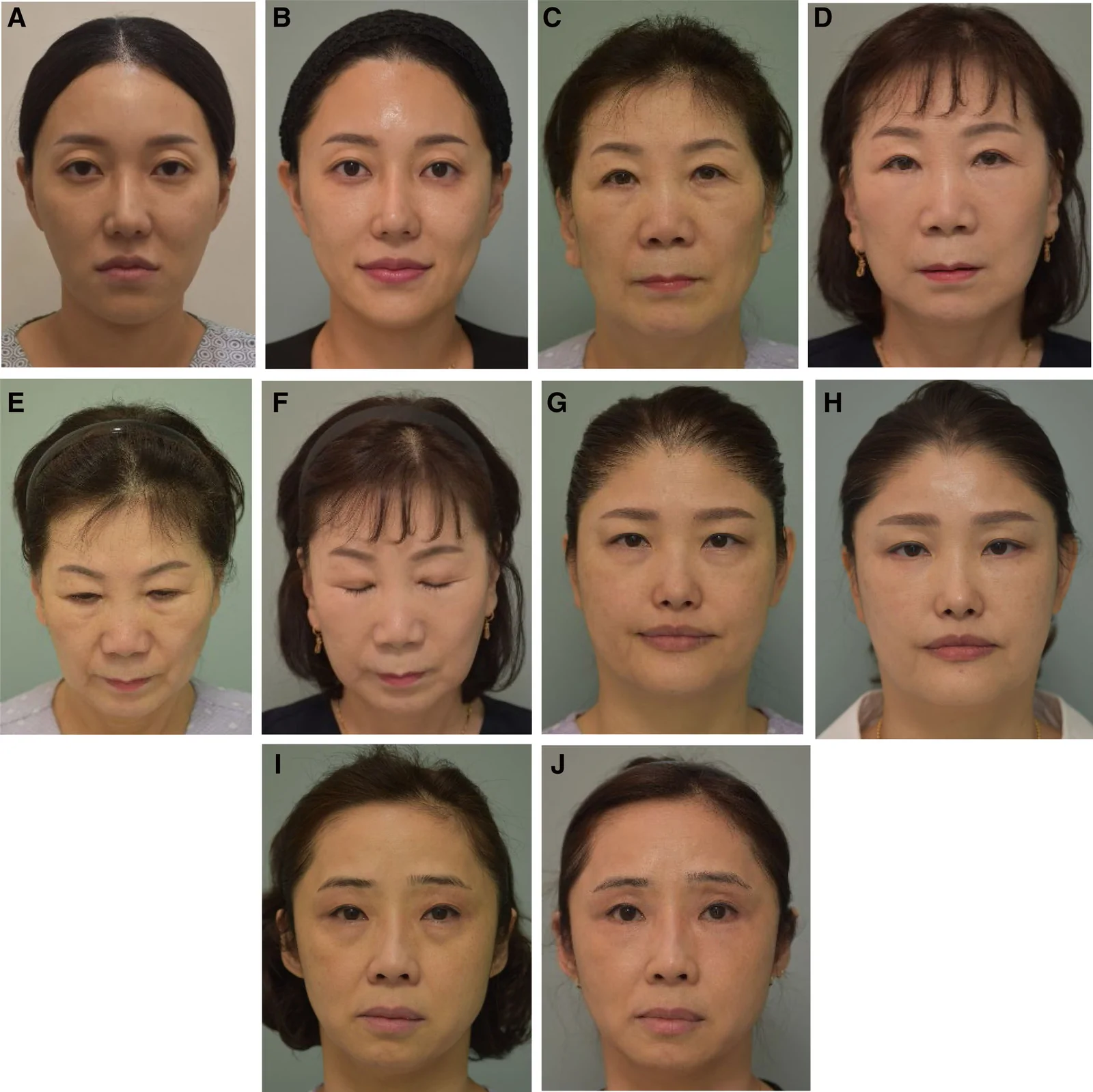
Fig. 5. Representative preoperative and postoperative photographs demonstrating correction of the lower eyelid asymmetry according to subtype-specific procedures. A, B, Type I-a—5 months postoperatively. Bilateral medial SOOF advancement and OOM suspension were performed, with an additional OOM suspension placed on the left side. C–F, Type I-b—61-year-old, 11 months postoperatively. Bilateral medial SOOF advancement and OOM suspension were performed, with an additional OOM suspension and lateral SOOF advancement on the left side. G and H, Type II-b—51-year-old who also underwent a concurrent forehead lift, 1 year postoperatively. Bilateral medial and lateral SOOF advancements and OOM suspension were performed, with an additional lateral SOOF advancement and additional OOM suspension on the left side. I and J Type III—49-year-old, 4 months postoperatively. Bilateral medial SOOF advancement and OOM suspension were performed, with two additional lateral SOOF advancements and an additional OOM suspension placed on the right side.各亚型术前术后照片:I-a(5 月,双侧内侧 SOOF 推进 + OOM 悬吊、左侧附加 OOM 悬吊)、I-b(61 岁,11 月)等,展示按亚型矫正的对称效果。No cases of lower eyelid retraction, ectropion, scleral show, infection, or clinically significant dry eye symptoms were observed during the follow-up period.
Systematic evaluation of lower eyelid asymmetry has received limited attention in plastic surgery literature. Much of the emphasis has been focused on indications and techniques, such as whether the lower blepharoplasty should be performed via the transconjunctival or subciliary approach. 19 , 20 Failure to recognize and address preexisting asymmetry invariably results in postoperative discrepancy. Symmetry of the lower eyelids is exceedingly uncommon, as differences in the size, vertical length, and spatial orientation of the maxilla and zygoma—the principal components of the inferior orbit—are nearly universal. 21 The etiology of such asymmetry is multifactorial, encompassing both genetic and environmental influences. 22
In our series of patients, the majority—96% (259 of 267)—demonstrated a posterior position of the left inferior orbital rim compared with the right. Additionally, a wider palpebro-malar groove was deemed noticeable on the left in 38% (102 of 267) of cases, whereas this pattern appeared on the right in only 2% (6 of 267). These findings reflect underlying skeletal discrepancies between the 2 zygomatic complexes.
Analysis of facial bone yawing, defined as rotation along the vertical axis, revealed that in 98% of cases, the right zygoma was positioned more anteriorly and superiorly, whereas the left zygoma was located more posteriorly and inferiorly. The larger facial volume on the left side compared to the right was a common finding in most individuals in our series. However, when the left facial skeleton rotates posteriorly, the left zygomatic complex may appear smaller on frontal view despite having greater actual volume, resulting in minimal apparent difference in palpebro-malar groove width. This asymmetric skeletal configuration exerts a direct influence on the overlying soft tissues. As the right facial skeleton tended to be smaller in volume, the smaller right zygoma predisposes the right midface to greater descent. However, its higher and more medial position results in a less prominent palpebro-malar groove in the lateral eyelid region and correspondingly less apparent lower eyelid sagging. Because soft tissues mirror the underlying osseous structure, lower blepharoplasty should incorporate asymmetric soft-tissue modification, as bony correction in this region is rarely performed. Procedures such as orbital fat repositioning, OOM suspension, and SOOF lift are important for rejuvenation of the lower eyelid and correction of tear trough deformity. 23 , 24
The palpebro-malar groove forms at the junction of orbital fat protrusion above the infraorbital rim and descent of the suborbicularis and malar soft tissues below, anchored by the orbicularis retaining ligament. Optimal correction, therefore, requires transposing orbital fat inferiorly to smooth the orbital fat bulge while elevating the descended midface to effectively efface the palpebro-malar groove. On the side with the wider palpebro-malar groove, broader redistribution of orbital fat is often performed; however, this approach alone is typically insufficient or inconsistent to correct asymmetry or improve the lid-cheek junction. 25 – 27 In our experience, the volume of orbital fat available for transposition is often inadequate to compensate for the underlying bony and soft-tissue discrepancy.
Other potential methods of correction, such as fat grafting, may compensate for soft-tissue deficiency in the palpebro-malar region; however, clinical outcomes are inconsistent, with reported resorption rates ranging from 20% to 80%. 28 An advantage of the SOOF utilization is its predictable viability, as it is a vascularized advancement flap and has not been shown to undergo resorption. Although soft-tissue descent on the left side may be less pronounced due to greater zygomatic volume, the caudally and posteriorly positioned zygoma compresses the tissue above the nasolabial fold, producing a puffy and aged appearance. Elevation of the left SOOF not only restores symmetry to the infraorbital region but also softens the nasolabial fold by re-elevating the midfacial descent.
On the side with greater lower eyelid vertical length, a single OOM suspension is often insufficient, as excessive tension leads to cheese-wiring. The orbicularis oculi is a thin, dynamic sphincter muscle composed of short, intrafascicularly terminating fibers without a broad aponeurotic sheet. 29 – 31 When a single suspension suture is placed directly into the OOM, the localized stress can exceed the tissue’s capacity to resist traction, resulting in progressive suture cut-through or cheese-wiring over time. We found that 2 fixation points are needed to provide additional support and offload force on each suture.
Insufficient advancement of the SOOF was the principal cause of suboptimal outcomes in our study. SOOF advancement—particularly in the lateral region—is critical for re-establishing symmetry. The SOOF is a fibrofatty layer located deep to the OOM and superficial to the periosteum of the zygoma and maxilla. 32 Reinforcement of the OOM suspension stabilizes the lateral contour and minimizes long-term relapse. Although concerns have been raised regarding lower eyelid retraction and dry eye symptoms following concurrent SOOF lift and lower blepharoplasty, 33 we did not encounter scleral show, ectropion, or dry eye syndrome in our series. We attribute this to secure fixation of the SOOF to the periosteum at the arcus marginalis, which avoids exerting a downward force on the lower lid. Moreover, meticulous hemostasis is critical, as cicatricial contracture from postoperative scarring is a significant contributor to lower eyelid retraction.
Previous authors have emphasized the importance of addressing midface descent during lower blepharoplasty. 26 In our experience, failure to correct lateral midface aging or asymmetry diminishes overall surgical satisfaction. Our utilization of SOOF repositioning in lower eyelid surgery addresses the medial midface ptosis. However, older patients often exhibit additional lateral midface descent; therefore, in cases with coexisting lateral midface ptosis, concurrent lateral midface lifting further enhances outcomes. Techniques such as minimally invasive cutaneous and SMAS suspension lift (CaSS) or traditional facelifts can be used to correct lateral midface ptosis. 34 , 35 Neglecting lateral midface ptosis often results in residual asymmetry, as the lower eyelid blends with the adjacent lateral cheek. Refinement of the eyelid-cheek junction is therefore key to achieving aesthetic coherence. 25 (Fig. 6 ).
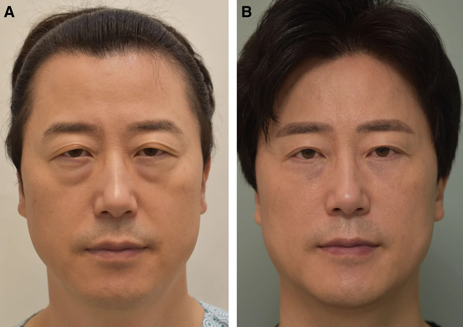
Fig. 6. Representative preoperative (A) and postoperative (B) photographs of a type II-a case, 2 years postoperatively. A 53-year-old patient underwent lower blepharoplasty, forehead lift, upper blepharoplasty, and minimally invasive lateral facelift.II-a 型(53 岁)术前(A)术后 2 年(B),联合下睑整形 + 额部提升 + 上睑整形 + 微创外侧提升,示重度伴中面部下垂者需联合外侧提升以获满意。Our results underscore the value of systematic, anatomy-based surgical technique rather than uniform bilateral approaches. Asymmetry is the norm rather than the exception; thus, detailed morphologic analysis should guide operative planning to achieve greater surgical refinement in lower blepharoplasty. Our algorithm is derived from our series and is intended as a framework for systematic planning and improvement of asymmetric lower blepharoplasty. Surgeons can consider the selective use of asymmetrical SOOF advancement and OOM suspension to address preexisting differences in the lower eyelid morphology.
The scope of the present study should be considered when interpreting these findings. The study reflects the experience of a single surgery center and lacks a comparative control group. Outcome assessment relied on standardized clinical photography, patient-reporting evaluation, and independent observer review, and follow-up duration was variable. Despite these considerations, the study provides a practical morphologic framework and algorithmic approach for addressing lower eyelid asymmetry in primary lower blepharoplasty.
Lower eyelid asymmetry is common and seems to be related to intrinsic differences in periocular skeletal anatomy. In this series, a morphologic classification based on palpebro-malar groove width, anterior-posterior displacement of the inferior orbital rim, and vertical length of the lower eyelid provided a structured framework for preoperative assessment. Application of an algorithmic approach incorporating SOOF advancement and OOM suspension was associated with improvement in lower eyelid symmetry in most patients. These findings support the value of systematic morphologic evaluation and tailored surgical planning when addressing lower eyelid asymmetry during primary lower blepharoplasty.
The authors have no financial interest to declare in relation to the content of this article.
Patients provided written consent for the use of their images.
术前评估要点(可直接迁移): 本文把下睑不对称拆成三个可测量维度——睑颊沟宽度、下眶缘前后位移、下睑垂直长度,并发现 96% 患者左下颌缘更后移、与颧骨复合体骨性不对称相关。对初次下睑整形(经皮肤或经结膜入路)的启示: 术前必须做『不对称形态学分析』,不能双侧对称式照搬方案 ;否则术后患者才『发现』原有不对称,易引发纠纷。我的下睑手术术前照相与触诊应常规记录这三参数。
SOOF 为何优于脂肪移植: 文中指出脂肪移植矫正睑颊沟软组织不足时吸收率 20%–80%、结果不稳定;而 SOOF 是带血管化推进瓣、可预测且不吸收。这与中面部提升中『用自身韧带 / 脂肪室复位而非填材料』的理念一致。算法把 SOOF 自内向外分 4 点固定(深鼻唇脂垫、内侧脂垫、外侧 SOOF、附加 SOOF),宽睑颊沟侧加做外侧段——是一套可复用的标准化手法。
OOM 悬吊的防切割细节: 下睑垂直长的一侧单悬吊易因局部应力过大导致『荷包线切割』(suture cut-through),本文用双固定点分散张力。这是实操中的关键防并发症点: 悬吊 OOM 时避免单点高张力,按需加第 2 个固定点 。外侧 SOOF 推进不足是效果欠佳的主因,提示术中『外侧中面部』不能被忽略。
循证边界: 单中心、无对照、随访时长不一(4–24 月)、结局依赖照片与评者主观。但样本量 267 例、94% 对称满意且无下睑退缩 / 外翻 / 干眼,证据强度对临床术式参考足够。对我团队的启发:下睑整形从『去脂 + 收紧』升级为『按骨性不对称做 SOOF 复位 + OOM 悬吊』的算法化规划,并把『外侧 SOOF 充分推进』列为质控点;重度伴外侧中面部下垂者联合外侧提升。
声明:中文精读 · 仅供学术参考。内容来自公开文献检索,不代表本人观点,不构成诊疗建议。 医疗美容需在正规医疗机构由执业医师实施。
基于相同主题推荐 · 继续深挖
背景:鼻唇沟(NLF)抚平是面部年轻化的难点之一,本述评分析各类除皱术式对 NLF 与中面部松弛长期矫正的证据。
背景:面部老化呈区域特异性凸度增加与沟槽空虚,需分层(浅脂肪层约 3.0 mm、SMAS 约 4.5 mm)而非均一能量;多数既往 HIFU 研究采用全脸均一参数,难兼顾相反容积向
背景:GLP-1 受体激动剂减重常伴随“Ozempic Face”式面部容积流失与皮肤松弛,其力学机制为张力稳态崩溃;透明质酸(HA)填充剂以占位为主、储存模量(G′)偏低,难以持