透明质酸填充剂与多核苷酸在眼周年轻化中的比较
2026年7月17日19 min read
Journal of Clinical Medicine · 2026;15(13):4971
Authors: Rabia S. Khan, Kashif Hafeez
Affiliations: 1: ICE Postgraduate Dental Institute and Hospital, 24 Furness Quay, Salford M50 3XZ, UK; 2: Dental Faculty Oral & Craniofacial Sciences, King's College London, London SE1 9SP, UK
Journal: Journal of Clinical Medicine | 2026-6-26 | PMCID: PMC13361502
DOI: 10.3390/jcm15134971
The periorbital region represents one of the most challenging anatomical sites in aesthetic medicine due to its thin dermis, complex vascularity, and susceptibility to oedema and contour irregularities. While hyaluronic acid (HA) fillers remain the gold standard for volumetric correction, their limitations in skin quality enhancement and risk of complications such as Tyndall effect and malar oedema have driven interest in regenerative alternatives. Polynucleotides (PN), particularly polydeoxyribonucleotides (PDRN), have emerged as bioactive agents capable of promoting dermal remodelling, angiogenesis, and anti-inflammatory responses. This review critically evaluates current evidence comparing PN and HA in periorbital rejuvenation, integrating mechanistic insights, clinical outcomes, and safety considerations. While HA remains superior for structural correction, PN demonstrates consistent improvements in dermal quality parameters, including elasticity, hydration, and fine rhytids, with a favourable safety profile. However, heterogeneity in study design, product formulation, and outcome measures limits the ability to draw definitive conclusions. Future research should prioritise standardised protocols, long-term follow-up, and direct comparative trials to establish optimal treatment algorithms.
Keywords: periorbital rejuvenation; hyaluronic acid fillers; polynucleotides; polydeoxyribonucleotides (PDRN); tear trough deformity; dermal remodelling; skin quality; regenerative aesthetics
The periorbital region represents one of the most anatomically and functionally complex areas in aesthetic medicine, and consequently, one of the most challenging to treat predictably. Unlike other facial regions, the infraorbital skin is exceptionally thin, approximately 0.5 mm, and is underpinned by a highly vascularised and dynamic musculoskeletal framework. This unique anatomical configuration not only accelerates the visible manifestations of ageing, but also significantly constrains the therapeutic window for safe and effective intervention [1,2].
From a structural perspective, ageing of the periorbital region is not a singular process but rather a multifactorial interplay between dermal atrophy, ligamentous laxity, redistribution of fat compartments, and alterations in microcirculation. The attenuation of the orbicularis retaining ligament, combined with volume loss in the deep medial cheek fat and pseudo-herniation of orbital fat, contributes to the formation of the tear trough deformity. Simultaneously, dermal thinning and reduced collagen and elastin content result in increased translucency, thereby accentuating underlying vasculature and pigmentation [3,4]. These layered changes culminate in a complex clinical presentation characterised not only by contour irregularities but also by compromised skin quality.
Additional anatomical structures play a critical role in periorbital ageing and treatment outcomes. The orbicularis retaining ligament and tear trough ligament contribute to the characteristic lid-cheek junction deformity through progressive attenuation and tethering. Furthermore, age-related changes in superficial and deep fat compartments, particularly the deep medial cheek fat and sub-orbicularis oculi fat (SOOF), contribute substantially to contour irregularities. The lymphatic drainage network of the lower eyelid and midface is equally important, as disruption or compression of these pathways may predispose patients to persistent oedema following injectable treatments.
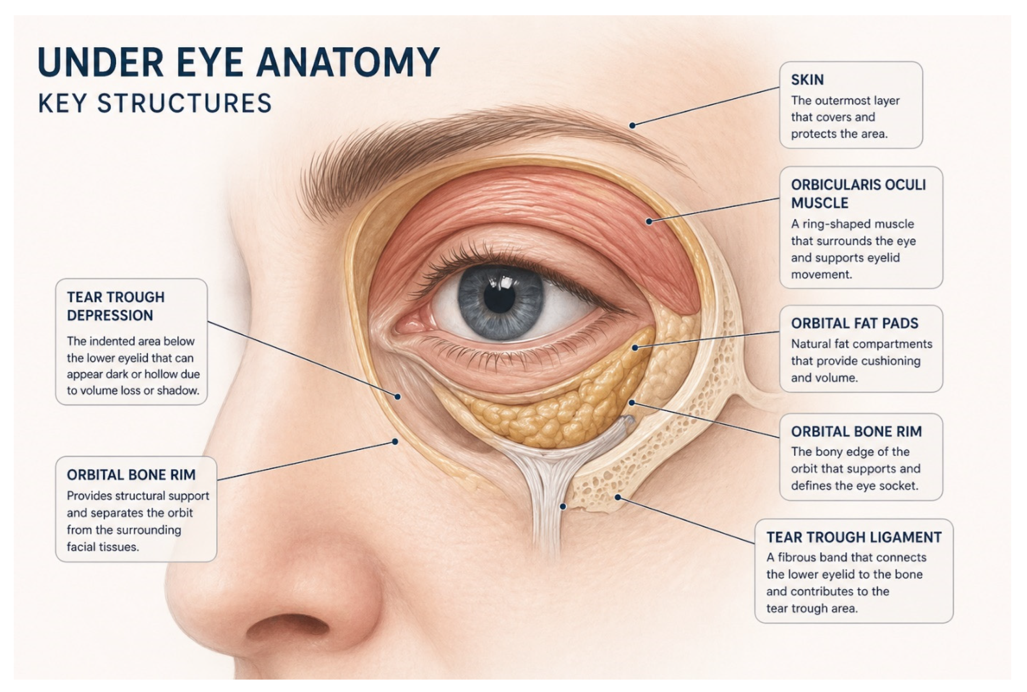
Figure 1 Under-eye (periorbital) anatomical structures, such as orbicularis retaining ligament, tear trough ligament, SOOF, deep medial cheek fat relevant to tear trough deformity and aesthetic treatment.
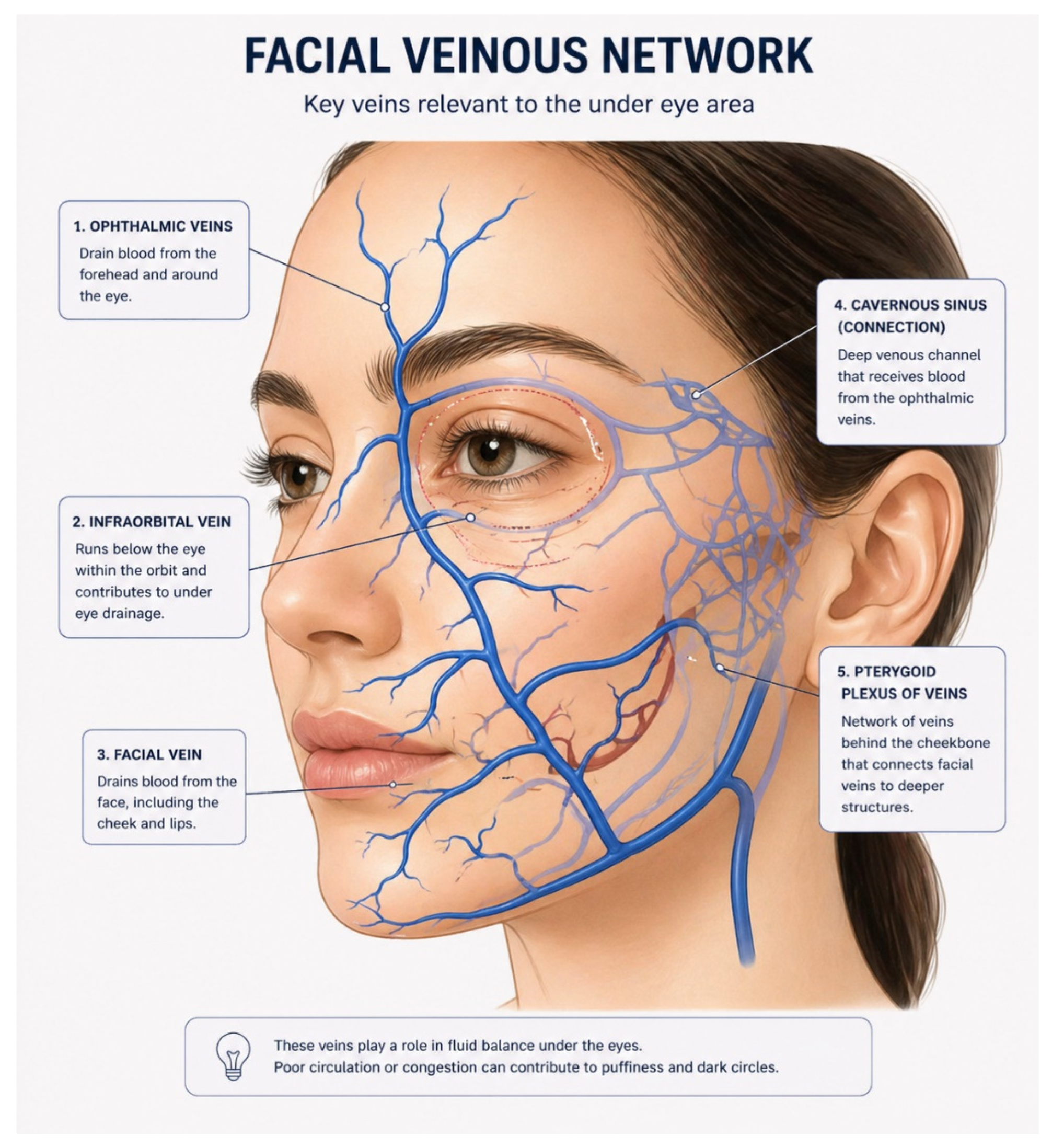
Figure 2 Facial venous anatomy relevant to the periorbital (under-eye) region.
Critically, this anatomical and physiological fragility renders the periorbital region highly susceptible to treatment-related complications. The dense vascular network and limited lymphatic drainage predispose patients to prolonged oedema, while the thin dermis increases the risk of visible filler placement and the Tyndall effect when using hyaluronic acid. Furthermore, the constant activity of the orbicularis oculi muscle introduces an additional dynamic component, whereby repetitive motion can influence product distribution, longevity, and aesthetic outcomes [4].
This complexity challenges the traditional paradigm of volumetric correction as a primary strategy for periorbital rejuvenation. While hyaluronic acid fillers have demonstrated efficacy in restoring contour deficits, their mechanism of action predominantly based on space occupation and water attraction, does not address the underlying dermal degeneration that contributes to fine rhytids, crepiness, and textural decline. In some cases, volumisation alone may exacerbate aesthetic concerns, particularly in patients with minimal volume loss but significant skin laxity or oedema-prone anatomy [5,6].
Therefore, a critical re-evaluation of treatment approaches in this region is warranted. Contemporary evidence increasingly supports a shift towards regenerative and skin-quality-focused interventions that target the underlying biological processes of ageing rather than solely its structural manifestations. Within this evolving framework, the anatomical complexity of the periorbital region should not be viewed merely as a limitation, but as a determinant guiding more nuanced, multimodal treatment strategies that balance structural correction with dermal regeneration [7,8].
This narrative review was undertaken to evaluate the current evidence regarding the use of hyaluronic acid (HA) fillers and polynucleotides (PN) for periorbital rejuvenation. A structured literature search was conducted using PubMed/MEDLINE, Scopus, Web of Science, and Google Scholar databases to identify relevant publications published between January 2015 and March 2026. The search combined Medical Subject Headings (MeSH) and free-text terms including: "periorbital rejuvenation," "under-eye rejuvenation," "tear trough," "hyaluronic acid filler," "dermal filler," "polynucleotide," "polydeoxyribonucleotide," "PDRN," "skin quality," "regenerative aesthetics," and "periocular rejuvenation."
Articles were screened for relevance based on title and abstract, followed by full-text review where appropriate. Priority was given to clinical trials, randomised controlled studies, systematic reviews, meta-analyses, consensus statements, and observational studies evaluating clinical outcomes, safety profiles, mechanisms of action, treatment protocols, or patient satisfaction associated with HA fillers and PN therapies in the periorbital region.
Studies were included if they:
Given the heterogeneity of study designs, treatment protocols, outcome measures, and PN formulations, a quantitative meta-analysis was not considered appropriate. Findings were therefore synthesised narratively.
Hyaluronic acid (HA) fillers have long been regarded as the cornerstone of minimally invasive periorbital rejuvenation, primarily due to their capacity to restore volume, their relative safety profile, and the availability of enzymatic reversal with hyaluronidase. Their widespread adoption is underpinned by predictable rheological properties, biocompatibility, and the immediate aesthetic improvement they provide in correcting contour deformities such as the tear trough [9,10].
The primary mechanism of HA fillers' space occupation combined with hydrophilic expansion renders them highly effective for structural augmentation. In the context of tear trough deformity, this allows for the restoration of the lid–cheek junction and improvement in shadowing. Nevertheless, this same hydrophilic property, which contributes to their volumising effect, also represents a fundamental drawback in the periorbital region. The propensity of HA to attract water can exacerbate fluid retention in an area already predisposed to lymphatic stasis, thereby increasing the risk of persistent malar oedema [11,12].
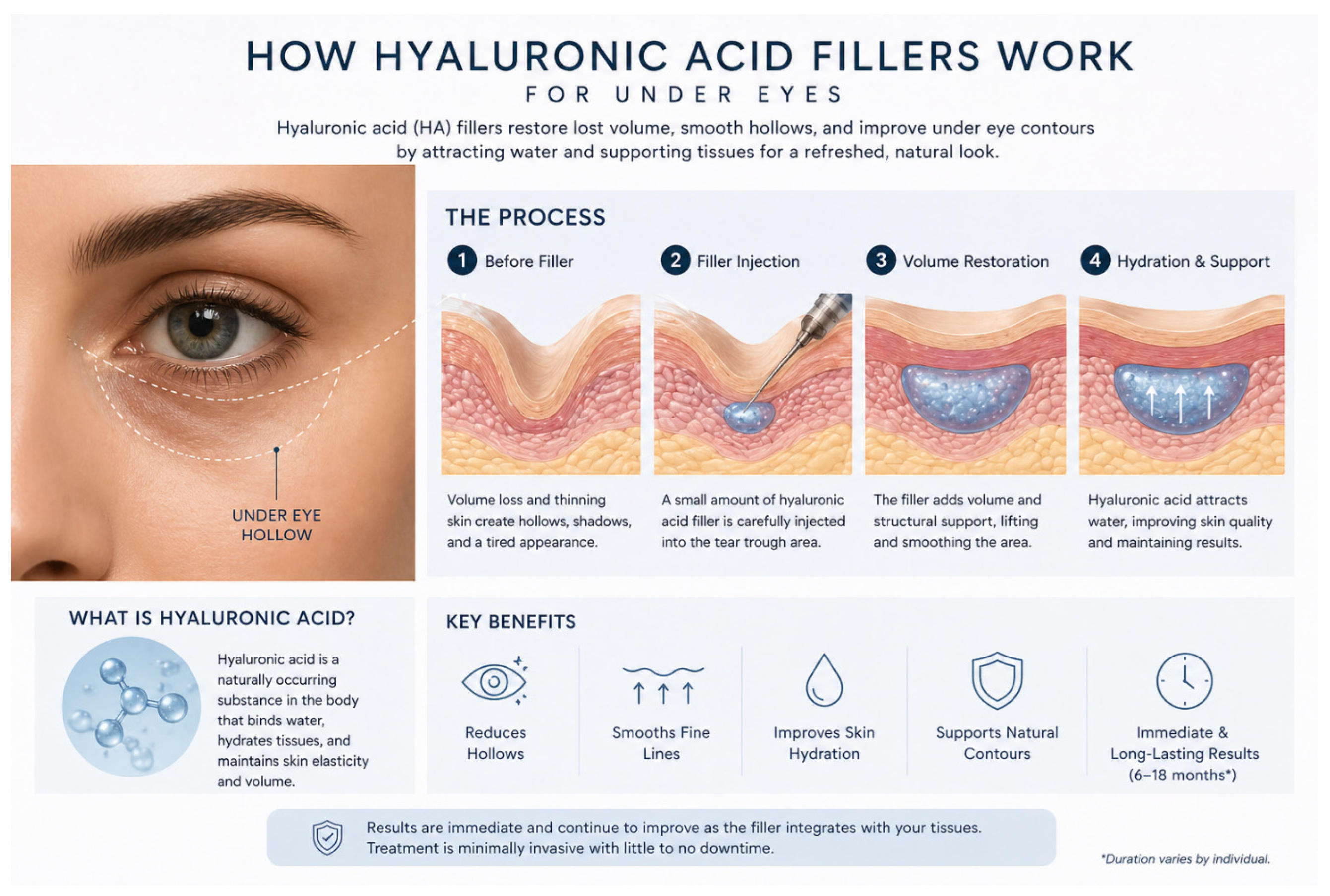
Figure 3 Mechanism of action of hyaluronic acid fillers in periorbital rejuvenation.
Furthermore, the thin dermal architecture of the periorbital skin amplifies the visibility of the superficially placed filler. The Tyndall effect, characterised by bluish discolouration due to light scattering, remains a well-documented complication and is largely a consequence of inappropriate depth of placement or unsuitable product selection [13,14].
Beyond aesthetic complications, the safety profile of HA fillers in the periorbital region warrants careful consideration. Although rare, vascular occlusion and subsequent visual compromise represent the most serious adverse events associated with filler injections. The proximity of the infraorbital and angular arterial systems, coupled with potential anastomoses with the ophthalmic circulation, necessitates a high level of anatomical knowledge and procedural caution [5,15].
Critically, HA fillers do not directly address the intrinsic changes associated with periorbital ageing at the dermal level. While they effectively correct volume deficits, they have limited impact on skin quality parameters such as elasticity, hydration balance, and fine textural irregularities. In patients where the primary concern is crepiness or dermal thinning rather than structural hollowing, volumisation alone may fail to achieve satisfactory outcomes [16].
Polynucleotides (PN), most commonly formulated as polydeoxyribonucleotides (PDRN), have emerged as a biologically driven alternative within aesthetic medicine, reflecting a broader paradigm shift from purely volumetric correction toward regenerative and tissue-modulating therapies [19,20]. Unlike hyaluronic acid fillers, which act predominantly through mechanical augmentation, PN exert their effects at a cellular and molecular level, targeting the fundamental processes underlying dermal ageing [21].
Mechanistically, PN function through activation of the A2A adenosine receptor, initiating a cascade of downstream effects that include fibroblast proliferation, increased synthesis of collagen types I and III, enhanced elastin production, and stimulation of angiogenesis. In parallel, PN engages nucleotide salvage pathways, facilitating cellular repair and regeneration while exerting anti-inflammatory effects [22,23].
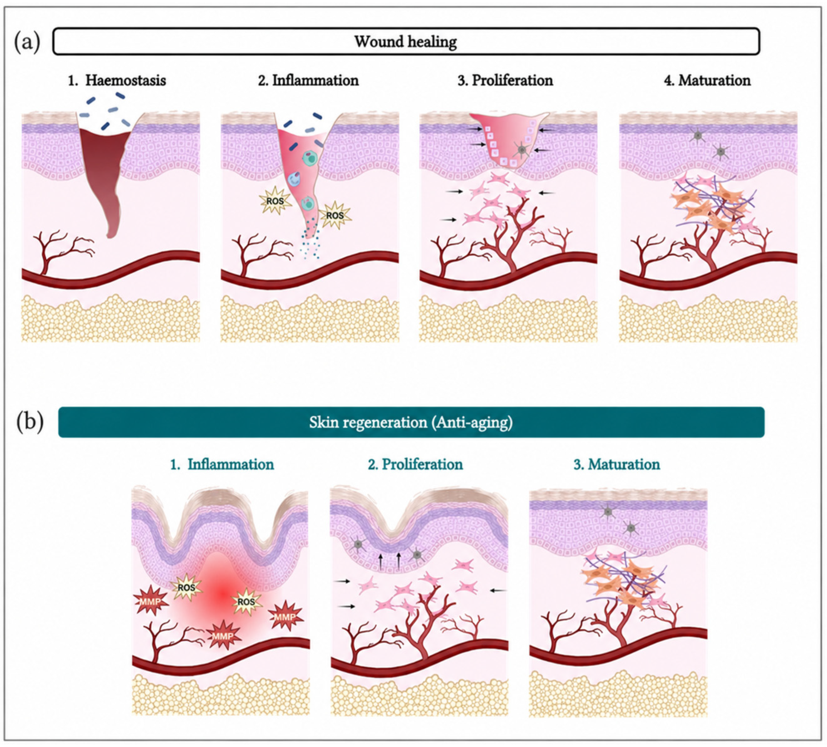
Figure 4 Comparison of classical wound healing and skin regeneration (anti-ageing) processes [3]. (a) Conventional wound healing progresses through four overlapping stages: haemostasis, inflammation, proliferation, and maturation. (b) Skin regeneration associated with anti-ageing therapies follows a similar but modulated pathway, primarily involving controlled inflammation, enhanced proliferation, and organised maturation, with reduced oxidative stress (ROS) and matrix degradation (MMPs).
Clinically, the application of PN in periorbital rejuvenation has been associated with improvements in fine rhytides, crepiness, and overall skin texture, often with a more subtle and progressive aesthetic outcome compared to HA fillers. This gradual onset of effect, while sometimes perceived as a limitation by patients seeking immediate results, may in fact represent a more naturalistic approach to rejuvenation, avoiding the risks of overcorrection and unnatural contour [24]. Importantly, the low volumetric impact of PN reduces the likelihood of complications associated with fluid retention, such as malar oedema [25].
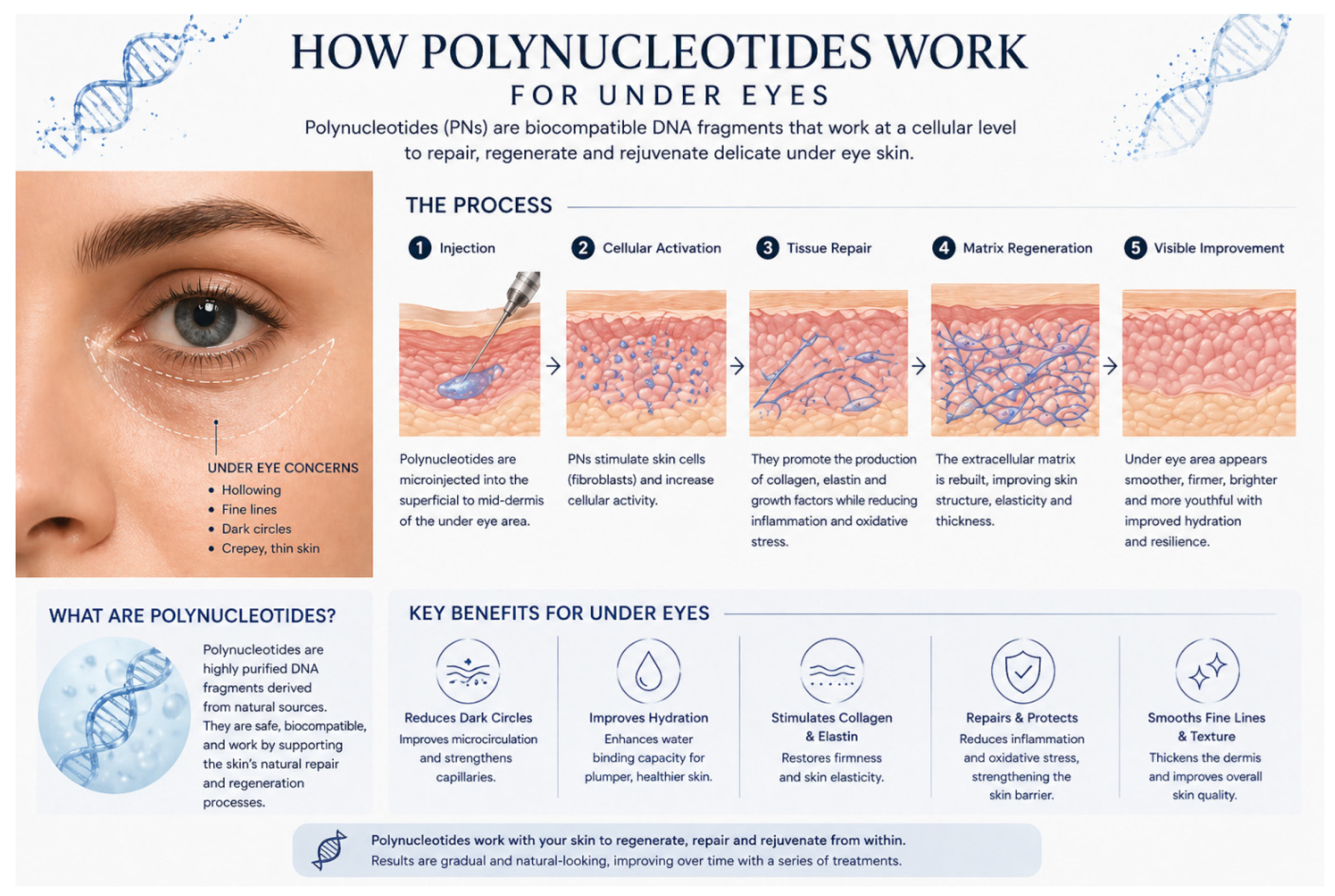
Figure 5 Mechanism of action of polynucleotides in periorbital rejuvenation.
However, despite these promising attributes, a critical evaluation of PN reveals several limitations. Firstly, the evidence base, although growing, remains relatively limited in scale and standardisation. Variability in PN formulations introduces heterogeneity that complicates direct comparison between studies [25]. Secondly, the magnitude of clinical improvement associated with PN, while statistically significant in many studies [26], is often modest when compared to the immediate and more pronounced effects achieved with HA fillers. PN do not provide structural support and therefore cannot address moderate to severe tear trough deformities or significant volume loss [26,27].
Lee et al., 2022 [30], evaluates PN compared with HA for periorbital rejuvenation. In a randomized, double-blind, split-face trial, PN fillers showed similar improvements in visual analogue and global aesthetic scores when compared with non-crosslinked HA. Notably, PN treatment demonstrated relatively greater improvements in skin elasticity, hydration, surface roughness, and pore volume over time. Both treatments were well tolerated, with no significant adverse events reported [30].
Table 1 GAIS and Response Trajectory (Illustrative HA vs. PN).
| Treatment Type | Typical GAIS/Aesthetic Response Pattern | Citations |
|---|---|---|
| Non-crosslinked/crosslinked HA fillers | High early GAIS responder rates (often >80–90%) from 1–3 months, sustained in many cases to 6–12+ months | [32,34,35,36] |
| PN monotherapy | Demonstrated wrinkle and hydration improvement over 4–6+ months; GAIS similar to HA in direct periocular comparison | [19,31] |
| PN–HA complexes | Case-level and mechanistic data suggest fast visible change plus regenerative skin-quality gains | [24,26] |
Evidence consistently shows that PN and HA differ more in safety profile than in efficacy, especially around the eyes. Across a systematic review of 219 PN-treated patients, side effects were generally mild and transient, with no serious complications reported [25]. Periorbital PN case series showed only mild swelling, discomfort, minor bruising at 2 days, and no delayed/persistent events such as nodules, prolonged oedema, Tyndall-like discoloration, or infection up to 6 months [38].
Reviews of periocular HA stress that most complications are also immediate, mild injection-related reactions. However, persistent or late malar/lower-eyelid oedema, blue discoloration (Tyndall effect), contour irregularities, filler migration, and chronic oedema are well documented and may appear weeks to years later [39,40,41,42]. Long-term follow-up of 147 patients found malar oedema in 11%, blue-grey dyschromia (Tyndall-like) in 31.3%, and contour irregularities in 30.5% [41].
Evidence supports PN as a biologically active, regenerative agent, but clinical facial outcomes are generally moderate and slow-onset, and not directly comparable to the rapid volumizing effects of HA fillers. PN and PN–HA formulations stimulate fibroblast proliferation, collagen production, ECM remodelling, angiogenesis, and migration in multiple in-vitro and animal models [24,39,46].
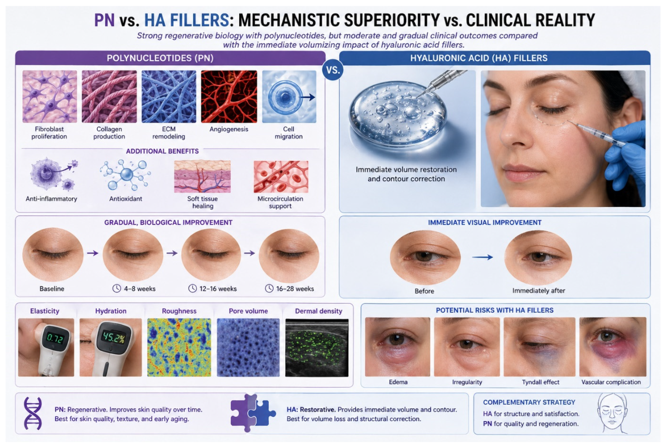
Figure 6 Mechanistic superiority versus clinical reality: polynucleotides versus hyaluronic acid fillers in periorbital rejuvenation.
Evidence supports using PN and HA as complementary, not competing, tools, with choice and sequencing tailored to the dominant clinical problem. HA remains the workhorse for groove/hollow correction. PN is repeatedly used as a skin booster for fine infraorbital/periorbital lines and texture.
A consensus on the PN-HPT priming paradigm recommends 2–4 PN sessions before fillers to recondition dermis and stabilize filler outcomes [55,56]. PN consensus guidance suggests face/periocular protocols of 3–4 sessions 2–3-weekly, followed by spaced maintenance [42].
PN is favoured for delicate, oedema-prone periocular skin and for patients seeking subtle, natural improvements without added volume [42,57]. HA tear trough guidelines stress strict selection (skin quality, vector, oedema tendency), minimal volumes (≤0.5 mL/eye), midface support first, and readiness to reverse with hyaluronidase [24,56].
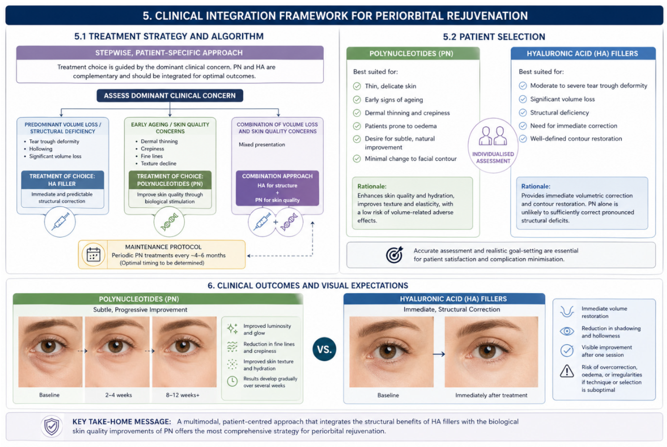
Figure 7 Clinical integration framework for periorbital rejuvenation using polynucleotides and hyaluronic acid fillers.
Current evidence aligns with a stepwise, patient-specific algorithm: HA fillers for clear volumetric/structural deficits, PN (alone or as PN-HA) for dermal quality and in oedema-prone or subtlety-seeking patients; and sequential PN + HA when both structure and tissue quality require treatment.
From a clinical perspective, three broad patient categories can be identified:
Interpretation of the current literature should be undertaken cautiously. Many PN studies involve small sample sizes, single-centre designs, and relatively short follow-up periods. Considerable heterogeneity exists between PN formulations with respect to molecular weight, concentration, purification methods, and treatment protocols. Outcome measures are also inconsistent. Furthermore, direct head-to-head comparative studies between PN and HA remain scarce, and long-term comparative data extending beyond 12 months are largely unavailable.
Current studies on PN and HA in periorbital and facial rejuvenation are promising but are small, heterogeneous, and often exploratory. Standardisation of PN formulations and treatment protocols is essential. Long-term follow-up studies extending beyond 12 months are particularly important. Direct comparative studies between PN and low G' hyaluronic acid fillers would provide valuable insights. Additionally, the integration of advanced imaging techniques and ultrasound-guided injections may enhance both safety and precision.
Polynucleotides represent a biologically driven advancement in periorbital rejuvenation, targeting key pathways involved in dermal ageing, including collagen synthesis, extracellular matrix remodelling, and tissue repair. Despite these mechanistic advantages, current clinical evidence indicates that their aesthetic effects are modest, gradual in onset, and not directly comparable to the immediate volumetric correction achieved with HA fillers. Rather than serving as a replacement, PN should be positioned as a complementary modality within a multimodal treatment framework. HA fillers remain the cornerstone for correcting volume loss and structural deficiencies, while PN contribute to improvements in skin quality, particularly in thin, delicate, or oedema-prone periorbital tissues. A critical synthesis of the available literature supports an integrated, patient-specific approach, in which structural augmentation and biological regeneration are strategically combined to optimise outcomes.
⚠️ 本文为医学文献全文精读,内容仅供学术参考,不构成医疗建议。
声明:中文精读 · 仅供学术参考。内容来自公开文献检索,不代表本人观点,不构成诊疗建议。 医疗美容需在正规医疗机构由执业医师实施。
基于相同主题推荐 · 继续深挖
2026/07/17 · 文献笔记 · 19 min read
HA填充剂在结构矫正方面优势明显,但存在水肿、Tyndall效应等风险;多核苷酸(PN/PDRN)在皮肤质量改善方面表现突出,包括弹性、水合度、细纹改善,安全性良好。两者应作为互补工具,HA用于结构性矫正,PN用于真皮再生。

系统综述表明,透明质酸(HA)填充剂在眶周结构性/容积矫正中仍占优势:早期GAIS应答率常达 80–90%(1–3个月),并多可维持至 6–12 个月以上。
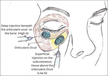
本研究提出双层差异G'值透明质酸填充注射技术,利用高G'值填充剂(骨膜上深层)提供结构支撑,低G'值填充剂(真皮下浅层)实现精细轮廓过渡,用于泪沟畸形矫正。5例病例系列显示高满意度(4.24/5分),效果可维持超过18个月,无严重并发症。该技术通过绕开中层血管危险区,安全性显著提升。