促红细胞生成素短期体外预处理增强纳米脂肪血管化与再生能力
2026年7月16日19 min read
Journal of Translational Medicine · 2026;24
Authors: Valeria Pruzzo, Francesca Bonomi, Ettore Limido, Andrea Weinzierl, Yves Harder, Matthias W. Laschke
Journal: Journal of Translational Medicine | 2026;24 | PMCID: PMC13366966 | PMID: 42443931
DOI: 10.1186/s12967-026-08640-x
Erythropoietin (EPO) is a glycoprotein hormone that exerts pro-angiogenic and anti-inflammatory effects. The present study investigated whether this beneficial profile of action is suitable for improving the in vivo performance of nanofat, an emulsified fat derivative that is clinically used in plastic and reconstructive surgery.
Repeated intravital fluorescent microscopic analyses showed that EPO-pretreated nanofat significantly accelerates and enhances the vascularization of the implants, as evidenced by an earlier onset of blood perfusion and an increased functional microvessel density when compared to controls. This was associated with a reduced inflammatory response to the implants, as indicated by lower numbers of adherent leukocytes in venules of the host tissue. Histological and immunohistochemical analyses further revealed an improved implant integration with an increased collagen I deposition and a higher density of nanofat-derived CD31+/green fluorescent protein (GFP+) microvessels, along with a reduced macrophage and neutrophil infiltration.
Nanofat was mechanically generated from subcutaneous adipose tissue of GFP+ C57BL/6J mice and incubated for 1 h in Hank's Balanced Salt Solution with or without EPO (3 IU/mL). The pretreated nanofat was seeded onto dermal substitutes, which were implanted into dorsal skinfold chambers of GFP- C57BL/6J mice and analyzed over 14 days.
These findings identify short-term pretreatment with EPO as an effective strategy to boost the vascularization and regenerative capacity of nanofat.
Erythropoietin (EPO) is a glycoprotein hormone that, beyond its hematopoietic function, exerts broad tissue-protective and regenerative effects, including the stimulation of angiogenesis as well as the suppression of apoptosis and inflammation [1,3,4,5].
Nanofat is another adipose tissue derivative already in clinical use for the treatment of chronic wounds and scars as well as for facial rejuvenation [6,9,10,12,13,14,15,16,17].
Based on the promising and broad application spectrum of EPO in plastic and reconstructive surgery and our own preliminary results on the ex vivo activation of nanofat, we analyzed in the present study, whether short-term exposure of nanofat to EPO (3 IU/mL) may effectively enhance its vascularization and regenerative capacity. For this purpose, EPO-pretreated and non-pretreated control nanofat was seeded onto dermal substitutes, which were analyzed in a well-established mouse dorsal skinfold chamber model by means of intravital fluorescence microscopy, histology and immunohistochemistry throughout an observation period of 2 weeks. Accordingly, it was possible to study the vascularization of the implants during the most critical initial phase after their implantation, which determines their barrier function and suitability for subsequent split-skin grafting.
Inguinal fat pads were harvested from 8 male and female green fluorescent protein (GFP)+ C57BL/6J donor mice.
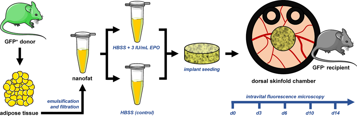
Fig. 1 Experimental protocol of the present study. Subcutaneous adipose tissue was harvested from GFP+ C57BL/6J donor mice and further processed by mechanical emulsification and filtration into nanofat, which was incubated for 1 h in HBSS with or without EPO (3 IU/mL). The pretreated nanofat was then seeded onto dermal substitutes, which were implanted into dorsal skinfold chambers of GFP- C57BL/6J recipient mice and repeatedly analyzed over 14 days by means of intravital fluorescence microscopy. Thereafter, the implants and their surrounding tissue were additionally examined by histology and immunohistochemistry.
All animal procedures were approved by the local authorities (permission number: 19-2024; State Office for Consumer Protection, Saarbrücken, Germany). The experiments were conducted in accordance with the European Directive 2010/63/EU on the protection of animals used for scientific purposes, the ARRIVE Guidelines and the National Institutes of Health (NIH) Guidelines for the Care and Use of Laboratory Animals (NIH publication #85−23 Rev. 1985).
For all surgical procedures and microscopic analyses, the mice were anesthetized by intraperitoneal injection of ketamine hydrochloride (100 mg/kg body weight; Ketabel®).
The white subcutaneous adipose tissue harvested from GFP+ donor mice was mechanically processed by emulsification and filtration to generate nanofat, which was then incubated for 1 h in Hank's Balanced Salt Solution (HBSS) with or without EPO (3 IU/mL).
The dermal substitute Integra® was seeded with either vehicle- or EPO-pretreated nanofat.
Dermal substitutes seeded with either vehicle- or EPO-pretreated nanofat were implanted into the observation window of dorsal skinfold chambers in GFP- recipient mice. This approach allowed longitudinal intravital fluorescence microscopy of the implants on days 0 (implantation), 3, 6, 10 and 14 (Fig. 1).
Eight regions of interest (ROIs) located at the border and center zones of the implants were analyzed for perfused ROIs (%) and functional microvessel density (cm/cm²).
The inflammatory response to the implants was assessed in four peri-implant postcapillary and collecting venules by analyzing microhemodynamic parameters (vessel diameter, centerline RBC velocity, shear rate and volumetric blood flow) and leukocyte-endothelial cell interactions. Leukocytes were classified as free-flowing, rolling or adherent.
After completion of the final intravital microscopy, the mice were euthanized by an overdose of anesthesia followed by cervical dislocation. The dorsal skin containing the implanted dermal substitutes was carefully excised and processed for histological and immunohistochemical stainings. Hematoxylin and eosin (HE) staining as well as immunohistochemical detection of CD31, collagen (Col) I, Col III, CD3, CD68 and myeloperoxidase (MPO) were performed.
The size of the individual groups (n) was based on previous experience and preliminary results. All data were analyzed using appropriate statistical tests, with p < 0.05 considered statistically significant.
Repeated intravital fluorescence microscopy enabled the detailed evaluation of the onset of blood perfusion and the progressive development of new microvascular networks in nanofat-seeded dermal substitutes. Notably, implants seeded with EPO-pretreated nanofat exhibited a much more pronounced microvascular ingrowth, both in the border and center zones over time, when compared to controls (Fig. 2).
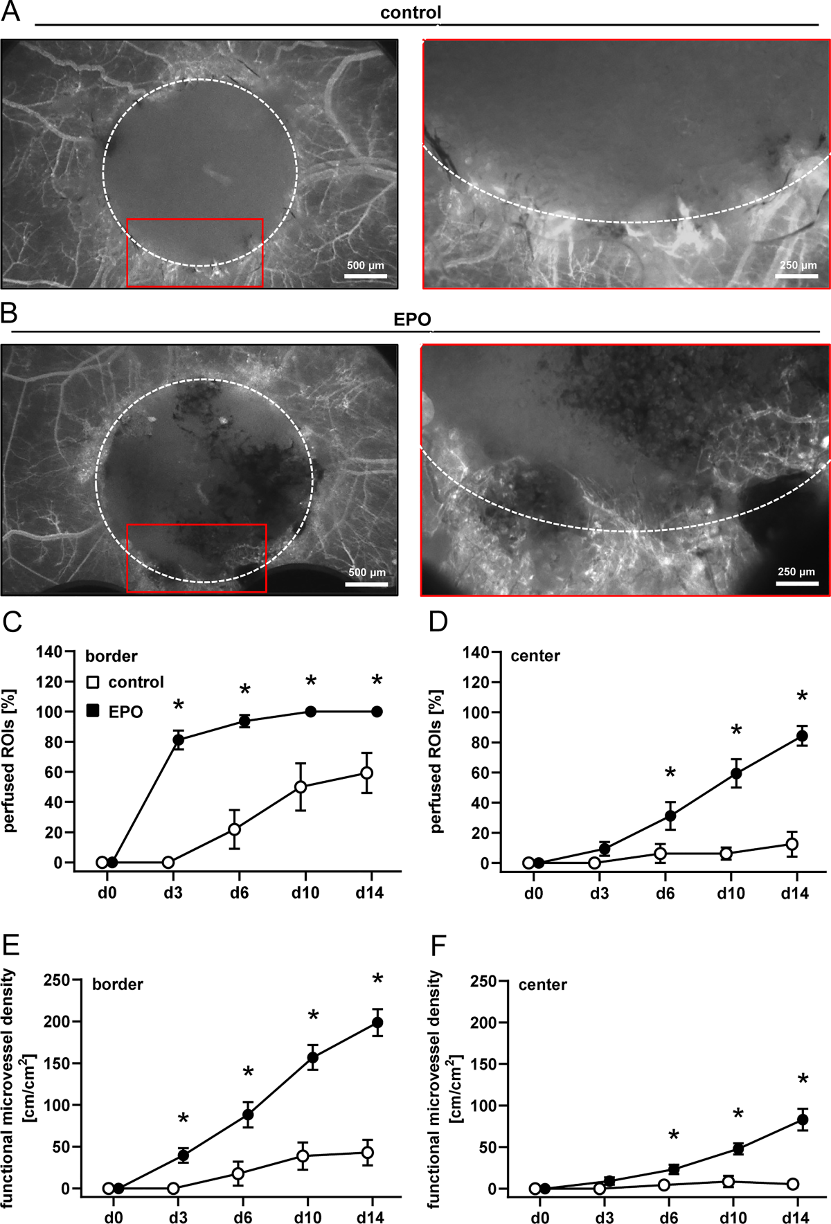
Fig. 2 In vivo microscopy of nanofat-seeded dermal substitutes. (A, B) Representative intravital fluorescence microscopic images of dermal substitutes seeded with vehicle-pretreated nanofat (control, (A)) and EPO-pretreated nanofat (EPO, (B)) on day 14 after implantation into dorsal skinfold chambers of C57BL/6J recipient mice (broken line indicates implant borders; red frame highlights regions of interest (ROIs) at the implant border shown in higher magnification on the right). (C-F) Perfused ROIs (%) (C, D) and functional microvessel density (cm/cm²) (E, F) in the border (C, E) and center zones (D, F) of dermal substitutes seeded with vehicle-pretreated nanofat (control; white circles, n = 8) and EPO-pretreated nanofat (EPO; black circles, n = 8), as analyzed by intravital fluorescence microscopy on day (d) 0, 3, 6, 10 and 14 after implantation. Mean ± SEM. *p < 0.05 vs. control.
Table 1 Diameter (µm), centerline RBC velocity (µm/s), shear rate (s−1) and volumetric blood flow (pL/s) of microvessels within the border and center zones of dermal substitutes seeded with vehicle-pretreated nanofat (control; n = 8) and EPO-pretreated nanofat (EPO; n = 8), as analyzed by intravital fluorescence microscopy on day (d) 0, 3, 6, 10 and 14 after implantation.
| Parameter | d0 | d3 | d6 | d10 | d14 |
|---|---|---|---|---|---|
| diameter (µm): | |||||
| border: control | - | - | 14.6 ± 0.0 | 15.4 ± 0.4 | 15.7 ± 1.0 |
| border: EPO | - | 12.2 ± 1.2 | 14.6 ± 0.9 | 14.2 ± 0.4 | 14.8 ± 0.3 |
| center: control | - | - | - | - | - |
| center: EPO | - | 9.0 ± 0.0 | 13.4 ± 1.7 | 13.4 ± 0.9 | 18.8 ± 3.6 |
To evaluate the inflammatory response to the implants, leukocyte-endothelial cell interactions were examined in peri-implant postcapillary and collecting venules of the surrounding subcutaneous host tissue (Fig. 3).
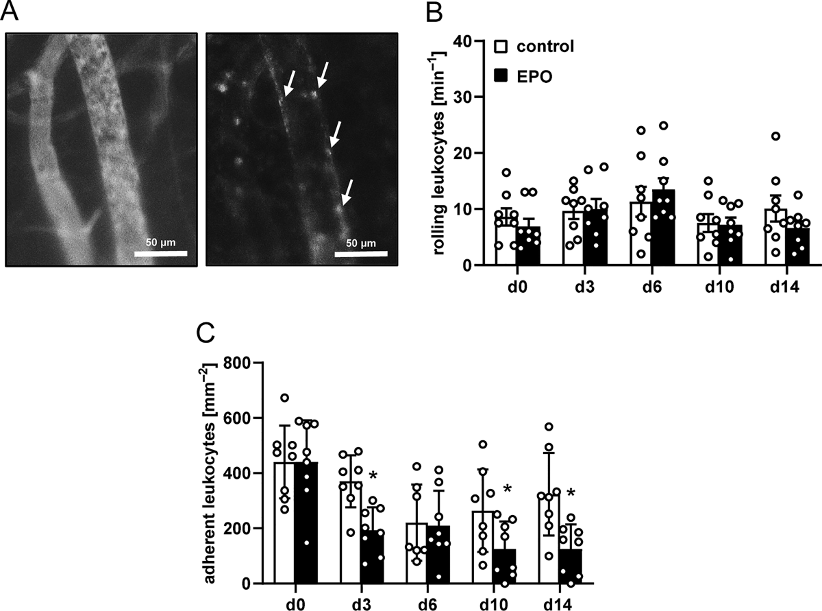
Fig. 3 Leukocyte-endothelial cell interaction in response to nanofat-seeded dermal substitutes. (A) Representative intravital fluorescence microscopic images of a collecting venule next to a dermal substitute seeded with vehicle-pretreated nanofat (blue light epi-illumination, contrast enhanced by 5% FITC-labeled dextran; left panel) and green light epi-illumination with in situ staining of leukocytes using 0.1% rhodamine 6G (right panel) (arrows indicate leukocytes). (B, C) Rolling leukocytes (min−1) (B) and adherent leukocytes (mm−2) (C) within postcapillary and collecting venules next to dermal substitutes seeded with vehicle-pretreated nanofat (control; white bars, n = 8) and EPO-pretreated nanofat (EPO; black bars, n = 8), as analyzed by intravital fluorescence microscopy on day (d) 0, 3, 6, 10 and 14 after implantation. Mean ± SEM. *p < 0.05 vs. control.
Table 2 Diameter (µm), centerline RBC velocity (µm/s), shear rate (s−1) and volumetric blood flow (pL/s) of postcapillary and collecting venules in direct vicinity to dermal substitutes seeded with vehicle-pretreated nanofat (control; n = 8) and EPO-pretreated nanofat (EPO; n = 8).
| Parameter | d0 | d3 | d6 | d10 | d14 |
|---|---|---|---|---|---|
| diameter (µm): | |||||
| control | 34.5 ± 0.8 | 34.7 ± 0.8 | 33.8 ± 0.0 | 32.3 ± 0.9 | 32.3 ± 0.8 |
| EPO | 34.5 ± 0.8 | 37.6 ± 1.2 | 35.2 ± 0.9 | 32.6 ± 1.2 | 32.6 ± 0.9 |
| centerline RBC velocity (µm/s): | |||||
| control | 303.9 ± 31.5 | 309.6 ± 26.7 | 312.7 ± 40.5 | 241.4 ± 25.8 | 293.5 ± 27.4 |
| EPO | 288.6 ± 24.3 | 266.7 ± 17.9 | 245.8 ± 15.0 | 235.7 ± 21.8 | 263.2 ± 17.6 |
| shear rate (s−1): | |||||
| control | 70.9 ± 7.1 | 71.4 ± 5.6 | 74.9 ± 8.7 | 60.0 ± 6.0 | 73.3 ± 6.6 |
| EPO | 67.4 ± 5.7 | 58.4 ± 4.2 | 55.8 ± 3.7 | 57.6 ± 5.0 | 64.1 ± 4.5 |
The implanted dermal substitutes were further examined by histology and immunohistochemistry at the end of the in vivo experiments on day 14. The analysis of HE-stained sections revealed an improved tissue integration of dermal substitutes seeded with EPO-pretreated nanofat when compared to controls (Fig. 4).
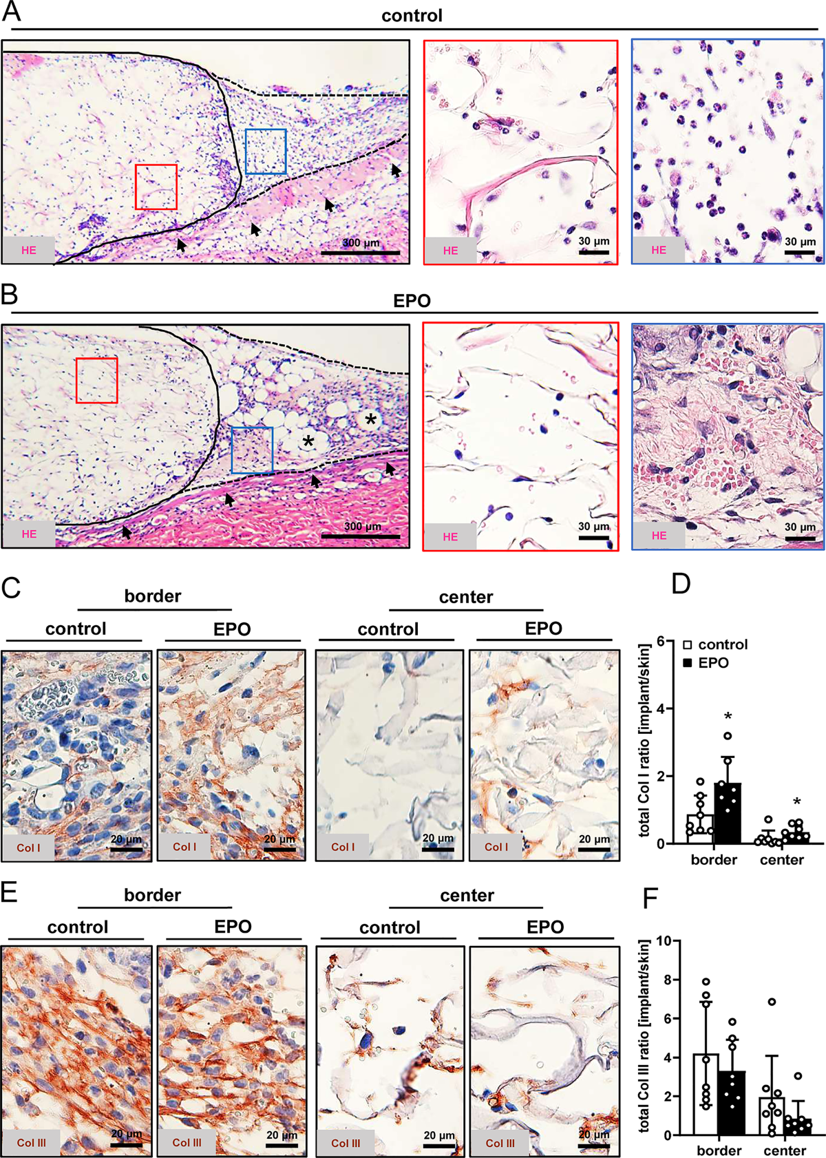
Fig. 4 Tissue integration of nanofat-seeded dermal substitutes. (A, B) Representative HE-stained sections of dermal substitutes seeded with vehicle-pretreated nanofat (control, (A)) and EPO-pretreated nanofat (EPO, (B)) on day 14 after implantation into dorsal skinfold chambers of C57BL/6J recipient mice (closed line indicates implant border; broken line indicates border zone; blue and red frames indicate ROIs in the border and center zones of the implants shown in higher magnification on the left panels; arrows indicate panniculus carnosus muscle; asterisks indicate adipocytes). (C, E) Representative immunohistochemical sections showing Col I (C) and III (E) in the border and center zones of dermal substitutes seeded with vehicle-pretreated nanofat (control) or EPO-pretreated nanofat on day 14. (D, F) Total Col I (D) and Col III (F) ratio (implant/skin) in the border and center zones of dermal substitutes seeded with vehicle-pretreated nanofat (control; white bars, n = 8) and EPO-pretreated nanofat (EPO; black bars, n = 8) on day 14, as analyzed by immunohistochemistry. Mean ± SEM. *p < 0.05 vs. control.
To further investigate tissue integration, the collagen content of the dermal substitutes was quantified, thereby distinguishing between Col I and Col III. This analysis revealed a significantly higher total Col I ratio in the border and center zones of dermal substitutes seeded with EPO-pretreated nanofat. In contrast, the overall Col III ratio did not differ between the two groups (Fig. 4).
Additional immunohistochemical analyses were performed to detect CD31+ microvessels (Fig. 5).
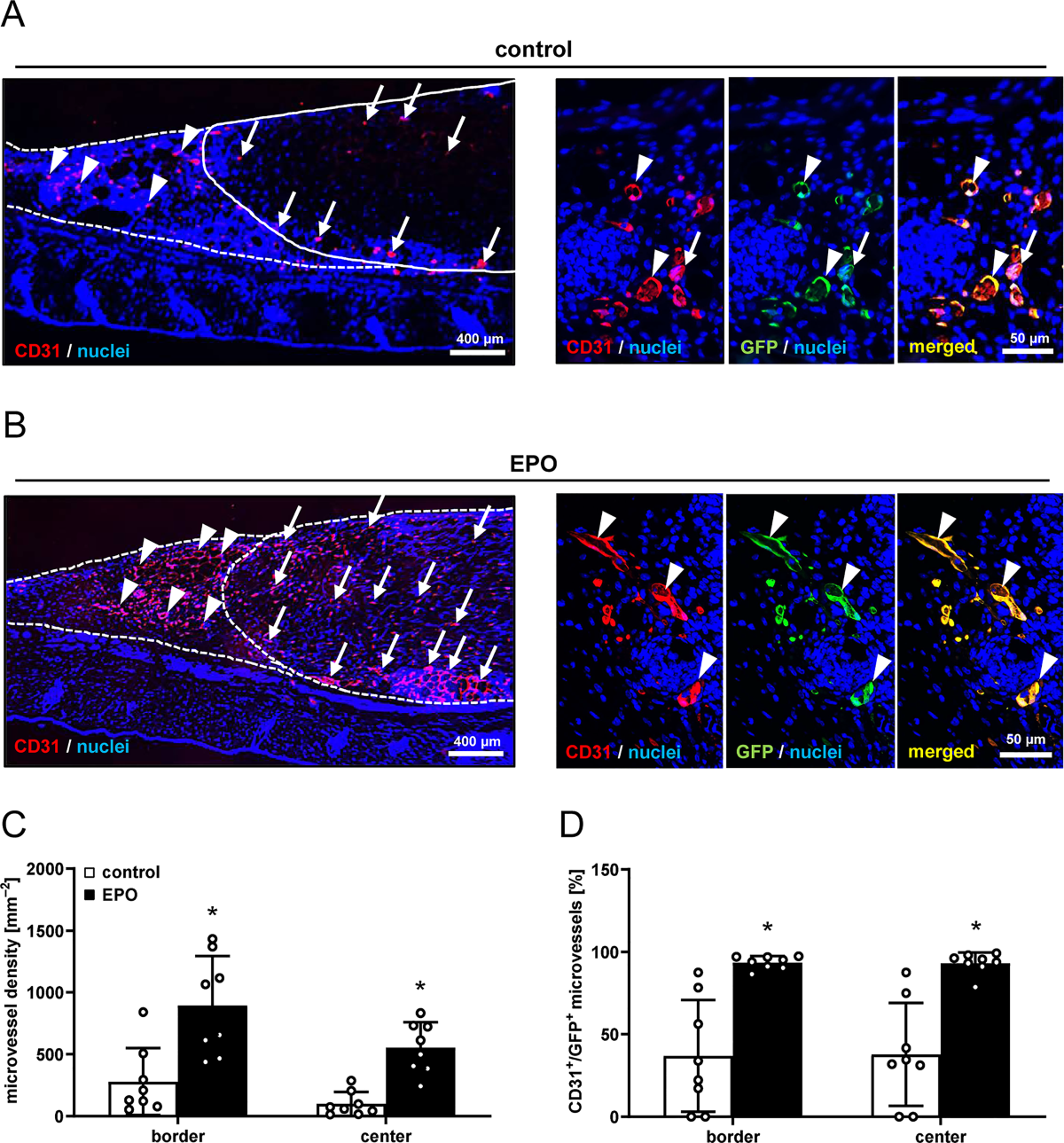
Fig. 5 Vascularization of nanofat-seeded dermal substitutes. (A, B) Representative immunohistochemical sections showing CD31+ microvessels in the border zones (arrowheads) and center zones (arrows) as well as the detection of CD31+/GFP− (arrows) and CD31+/GFP+ (arrowheads) microvessels of dermal substitutes seeded with vehicle-pretreated nanofat (control) or EPO-pretreated nanofat on day 14 after implantation into dorsal skinfold chambers of C57BL/6J recipient mice (closed line indicates implant border; broken line indicates border zones). (C) Microvessel density (mm−2) of dermal substitutes seeded with vehicle-pretreated nanofat (control; white bars, n = 8) and EPO-pretreated nanofat (EPO; black bars, n = 8) on day 14, as analyzed by immunohistochemistry. (D) CD31+/GFP+ microvessels (%) in the border zones and center zones of dermal substitutes seeded with vehicle-pretreated nanofat (control; white bars, n = 8) and EPO-pretreated nanofat (EPO; black bars, n = 8) on day 14, as analyzed by immunohistochemistry. Mean ± SEM. *p < 0.05 vs. control.
Finally, histological sections were stained with antibodies against CD3+, MPO+, and CD68+ cells (Fig. 6).
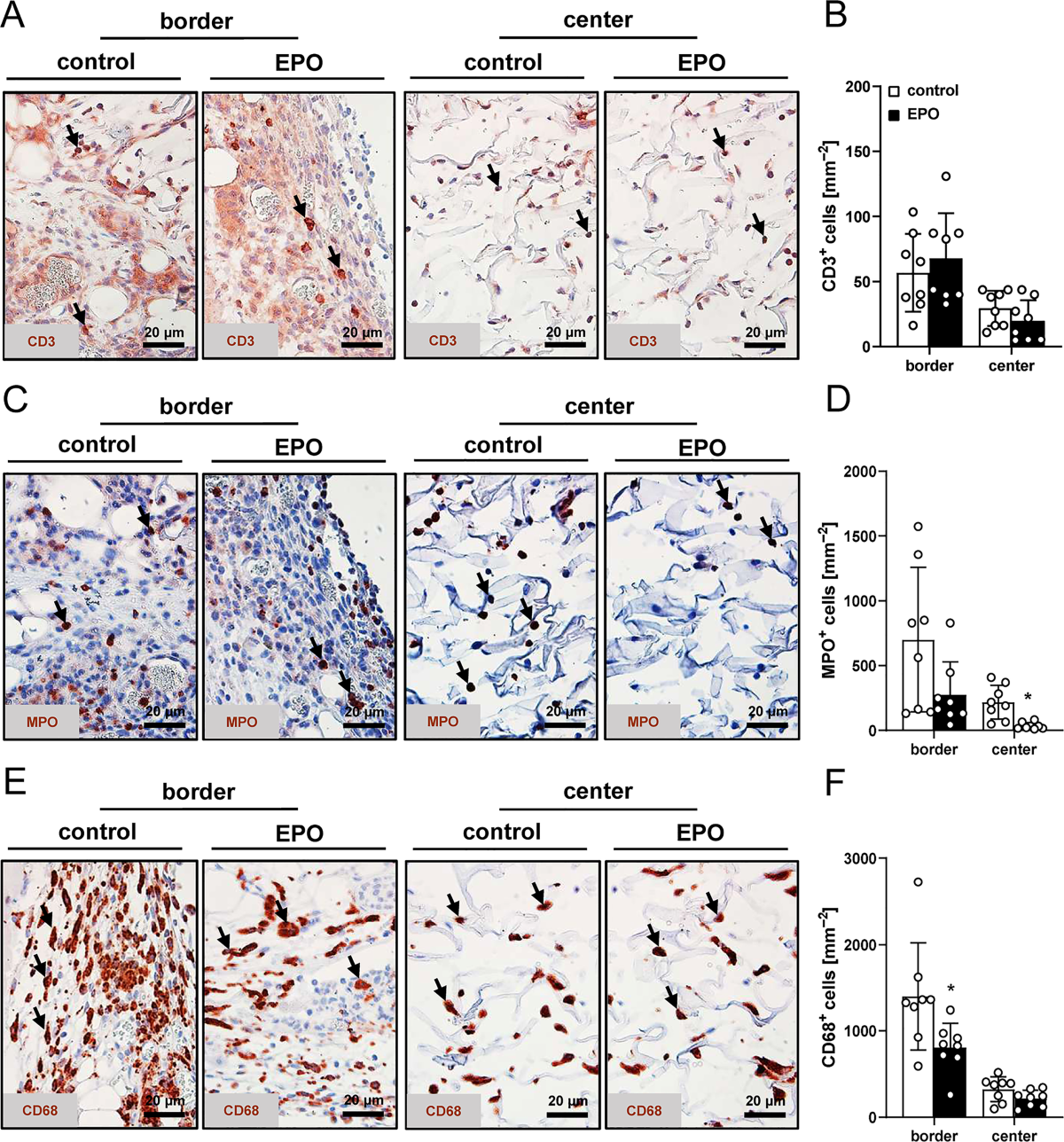
Fig. 6 Infiltration of immune cells into nanofat-seeded dermal substitutes. (A, C, E) Representative immunohistochemical sections showing CD3+ lymphocytes (A, arrows), MPO+ granulocytes (C, arrows) and CD68+ macrophages (E, arrows) in the border and center zones of dermal substitutes seeded with vehicle-pretreated nanofat (control) and EPO-pretreated nanofat (EPO) on day 14 after implantation into dorsal skinfold chambers of C57BL/6J recipient mice. (B, D, F) CD3+ lymphocytes (mm−2) (B), MPO+ granulocytes (mm−2) (D) and CD68+ macrophages (mm−2) (F) in the border and center zones of dermal substitutes seeded with vehicle-pretreated nanofat (control; white bars, n = 8) and EPO-pretreated nanofat (EPO; black bars, n = 8) on day 14, as analyzed by immunohistochemistry. Mean ± SEM. *p < 0.05 vs. control.
The regenerative capacity of nanofat can be further enhanced by exposure to bioactive stimuli, such as growth factors, platelet-rich fibrin, platelet-rich plasma, and glucose [17,19,23].
The vascularization capacity of nanofat was analyzed in the mouse dorsal skinfold chamber model by means of repeated intravital fluorescence microscopy. Dermal substitutes seeded with EPO-pretreated nanofat exhibited RBC-perfused microvessels as early as day 3 after implantation, whereas controls showed a delayed and spatially restricted perfusion. This early onset of vascularization was accompanied by a sustained increase in functional microvessel density over time, particularly within the central regions of the implants, which are typically most vulnerable to hypoxia [24].
Notably, the vessels located in the peri-implant tissue as well as within the border and center zones of the implanted dermal substitutes did not exhibit alterations in microhemodynamic parameters, including vessel diameter, centerline RBC velocity, shear rate or volumetric blood flow, throughout the 14-day observation period. Accordingly, EPO pretreatment did not induce abnormal vessel remodeling or hyperperfusion.
Besides its pro-angiogenic effects, EPO also significantly modulated the inflammatory response to the implants. While leukocyte rolling in peri-implant venules was comparable between the experimental groups, the number of firmly adherent leukocytes was consistently reduced in the group of dermal substitutes seeded with EPO-pretreated nanofat. Because leukocyte adhesion represents a critical step preceding transendothelial migration into tissues, these findings indicate a selective attenuation of excessive inflammatory cell recruitment rather than a global suppression of immune surveillance [28].
In line with this view, histological analyses on day 14 showed an improved tissue integration of dermal substitutes seeded with EPO-pretreated nanofat. Enhanced granulation tissue formation at the implant borders together with a higher number of residual adipocytes indicated a better survival of the transplanted adipose tissue components.
Immunohistochemical detection of CD31+ microvessels further demonstrated that EPO pretreatment significantly increases the microvessel density within the implants. Notably, more than 90% of these vessels were GFP+, indicating their origin from the seeded nanofat. On the other hand, only 37% of vessels in the control group were GFP+. These findings underscore the central function of EPO in promoting nanofat-dependent vascularization over host tissue-driven angiogenesis.
The present study further illustrates the enhanced biocompatibility of dermal substitutes seeded with EPO-pretreated nanofat. The significantly reduced density of CD68+ macrophages and MPO+ neutrophilic granulocytes within EPO-treated implants provides further evidence for an improved inflammatory microenvironment.
The choice of EPO concentration and exposure duration represents a critical aspect in EPO therapy. EPO exerts most of its properties in a dose-dependent manner [29,43].
Finally, some limitations of the present study should be considered. The experiments were conducted in a mouse dorsal skinfold chamber model, which allows high-resolution analysis of vascularization but does not fully recapitulate the complexity of clinical wound environments. Furthermore, this approach only enables the assessment of implant vascularization during the first 14 days. Future studies should address dose- or time-response relationships.
This study shows that short-term ex vivo pretreatment of nanofat with EPO is an effective and clinically translatable strategy to enhance vascularization, improve tissue integration and modulate inflammation in nanofat-seeded dermal substitutes. By accelerating the establishment of a functional microvascular network without compromising vessel quality or biocompatibility, this approach addresses a key limitation of dermal substitutes and holds promise for improving their future performance in reconstructive surgery and complex wound management.
⚠️ 本文为医学文献全文精读,内容仅供学术参考,不构成医疗建议。
声明:中文精读 · 仅供学术参考。内容来自公开文献检索,不代表本人观点,不构成诊疗建议。 医疗美容需在正规医疗机构由执业医师实施。
基于相同主题推荐 · 继续深挖
2026/07/16 · 文献笔记 · 19 min read
促红细胞生成素(EPO)短期体外预处理纳米脂肪可显著加速并增强植入物血管化,表现为更早的血液灌注和更高的功能性微血管密度,同时降低炎症反应、提高胶原蛋白I沉积和CD31+/GFP+微血管密度,减少巨噬细胞和中性粒细胞浸润。
鼻部塑形疗效与安全性:12个月前瞻性研究/PMC13100342_fig1.jpg)
这项单中心前瞻性研究纳入24名中国女性受试者,接受CaHA凝胶鼻部注射(骨膜上/骨膜浅层,单侧单次总量≤2.0 mL,平均0.93±0.22 mL),全部完成12个月随访,无脱落。
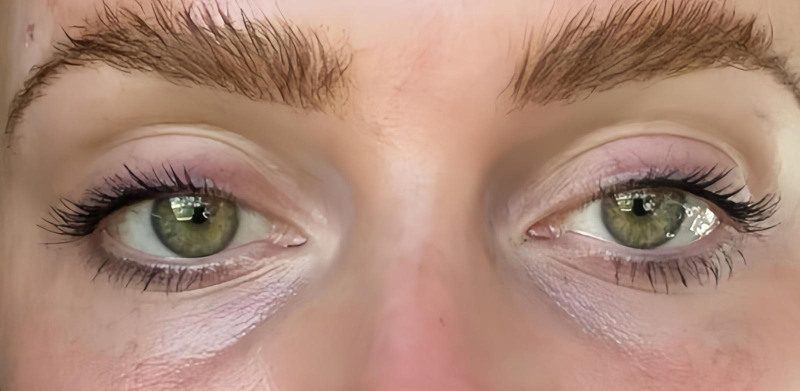
5 例健康女性(35–50 岁,中东与白人)接受 VAMP(多核苷酸基生物刺激剂,Prollenium)泪沟注射,每侧 1 mL(共 2 mL),23G 30mm 钝针,微滴 +